← epiphysis and articular cartilage Epiphysis labeled arteries cartilage epiphyses articular diaphysis shaft anatomyqa periosteum hyaline covered stippled arterial labeling layer ends called marrow portions ishowspeed duke dennis Duke dennis-ishowspeed "diss track" →
If you are looking for Proximal Phalanx Foot Anatomy you've came to the right web. We have 35 Images about Proximal Phalanx Foot Anatomy like Distal Phalangeal Joint, Distal Phalanx: Definition, Location, Anatomy, Diagram and also Phalanges Bones. Read more:
Proximal Phalanx Foot Anatomy
 mungfali.com
Distal Phalanx
 fity.club
Human Skeleton Anatomy, Human Body Anatomy, Muscle Anatomy, Hand Bone
 www.pinterest.com
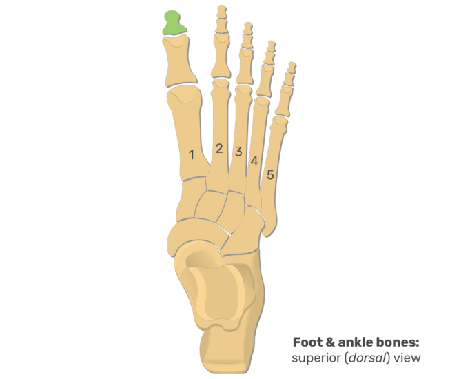
Metatarsal Bones And Foot Phalanges: Anatomy And Diagram | GetBodySmart
 www.getbodysmart.com
Distal Phalangeal Joint
 mavink.com
Foot Trauma, Fracture Of The Interphalangeal Joint Of The Great Toe
 cityfootcare.com
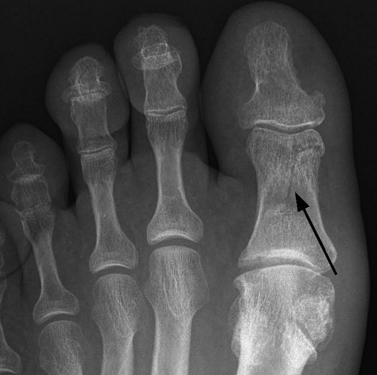
Atraumatic Fracture Of Proximal First Phalanx | CMAJ
 www.cmaj.ca
phalanx proximal fracture cmaj atraumatic
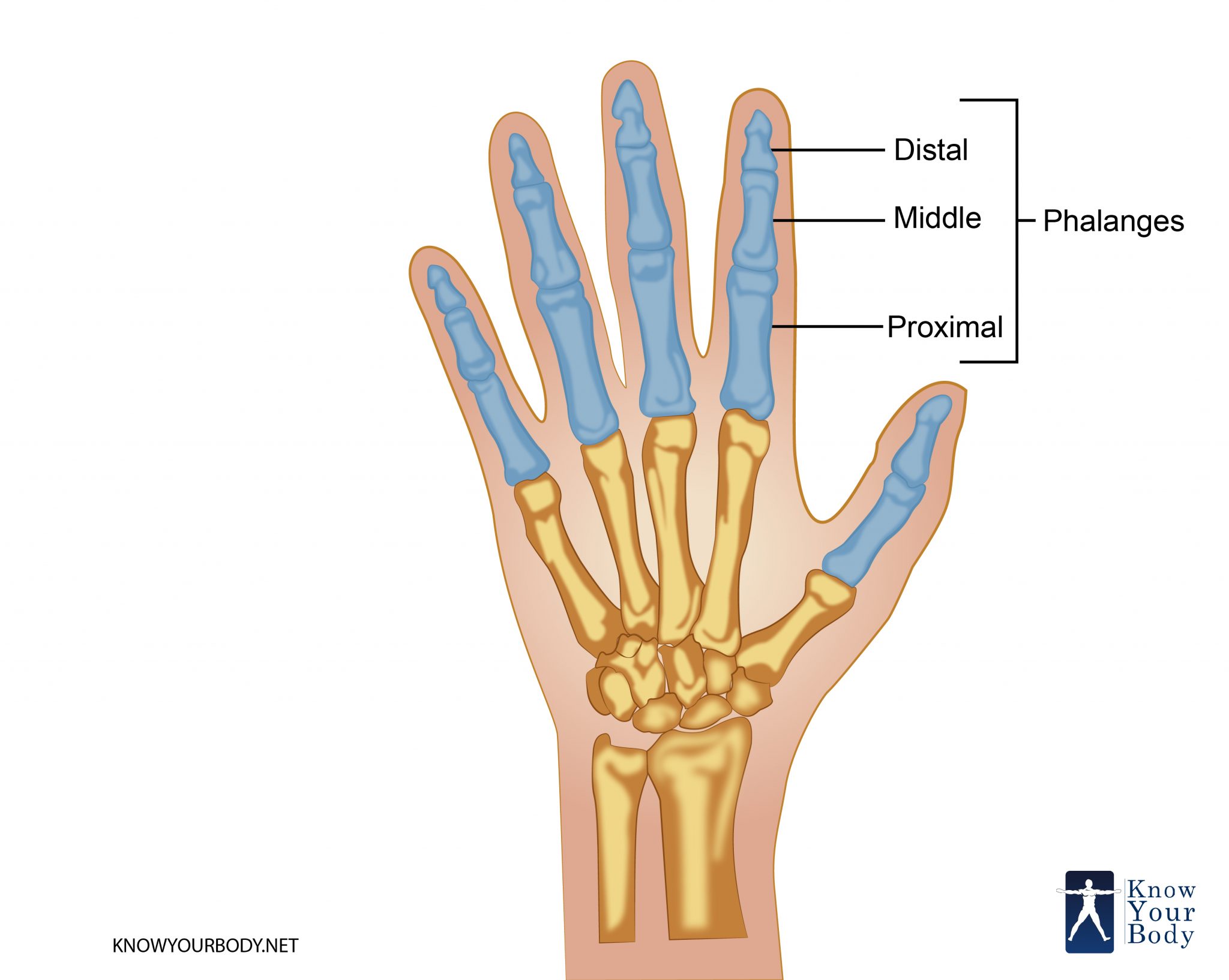
Phalanges Bones
 ar.inspiredpencil.com
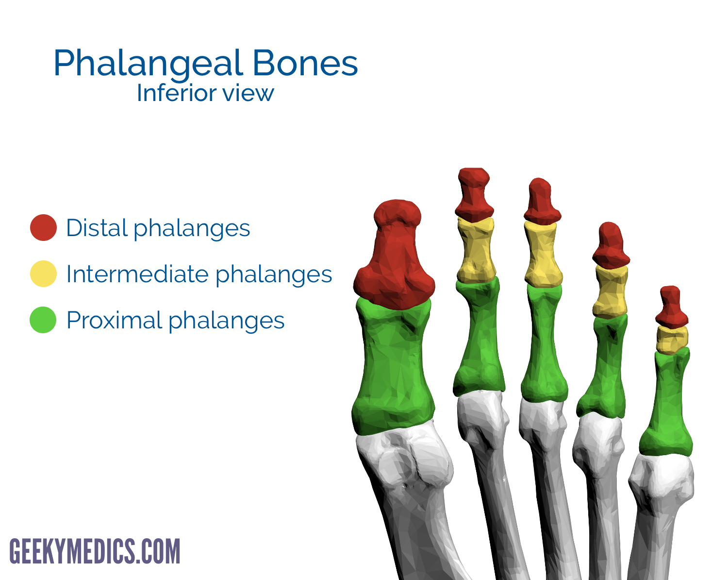
Proximal Phalanx Foot Anatomy
 mungfali.com
Cleft Epiphysis | Radiology Reference Article | Radiopaedia.org
 radiopaedia.org
epiphysis cleft phalanx proximal osteochondritis dissecans radiopaedia radiology ocd hallux joint
Cleft Epiphysis | Radiology Reference Article | Radiopaedia.org
 radiopaedia.org
epiphysis cleft proximal phalanx radiopaedia radiology
F2
 www.omjournal.org
[DIAGRAM] Proximal Epiphysis Long Bone Diagram - MYDIAGRAM.ONLINE
![[DIAGRAM] Proximal Epiphysis Long Bone Diagram - MYDIAGRAM.ONLINE](https://www.orthogate.org/press/wp-content/uploads/2017/09/MM-01-001-fig02.png) mydiagram.online
mydiagram.online
Distal Phalangeal Joint
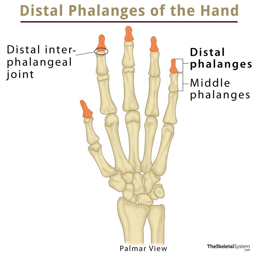 mavink.com
Hand Epiphyses Demonstrate Differences In Maturation On Posteroanterior
 www.researchgate.net
First Distal Phalanx (toe) Fracture | Image | Radiopaedia.org
 radiopaedia.org
Cureus | Type 2 Salter-Harris Physeal Injury Of The Proximal Phalanx Of
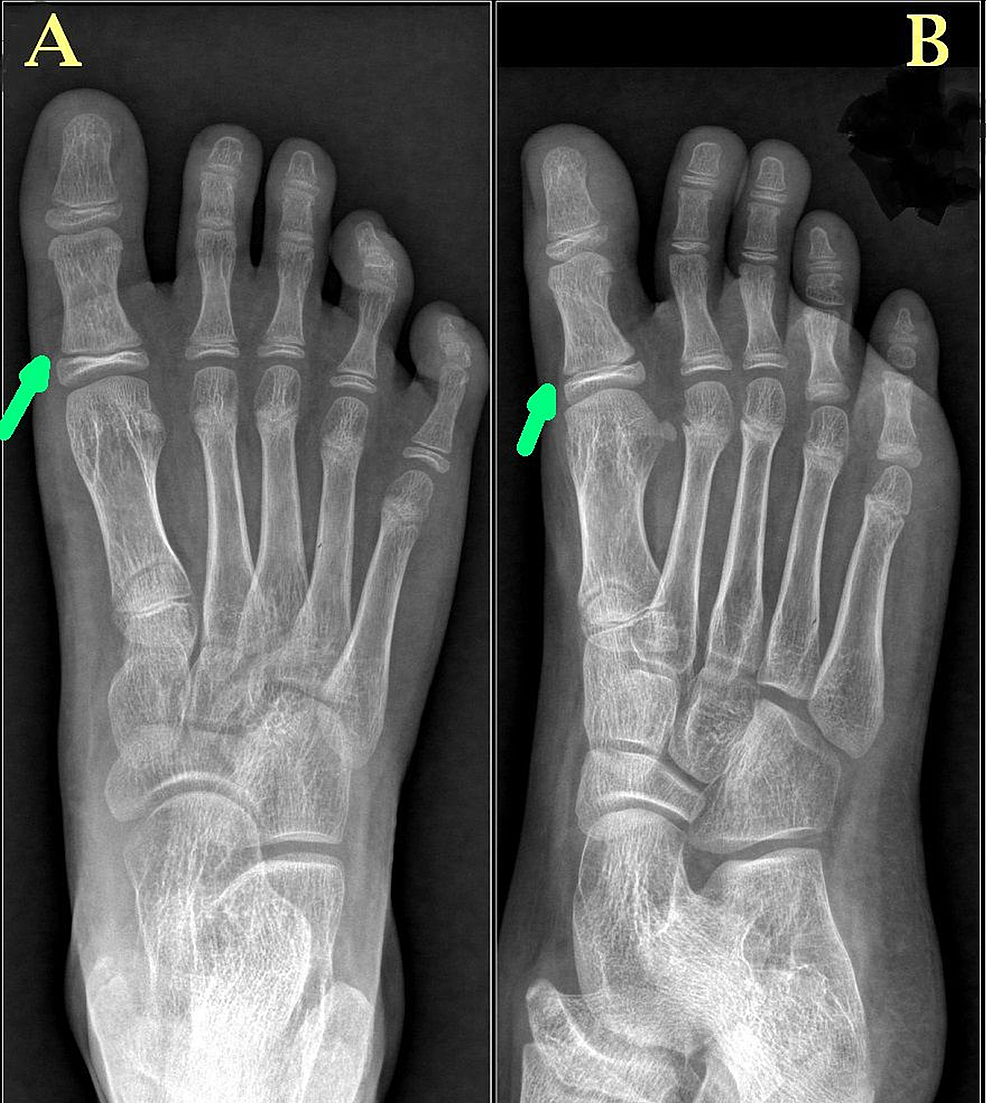 www.cureus.com
salter harris phalanx proximal oblique physeal radiographs anteroposterior
First Metatarsal Bone Location, Anatomy, & Diagram
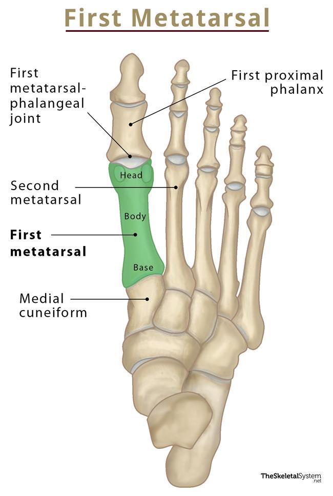 www.theskeletalsystem.net
Toe Bones (Phalanges Of The Foot) – Anatomy, Location, & Diagram
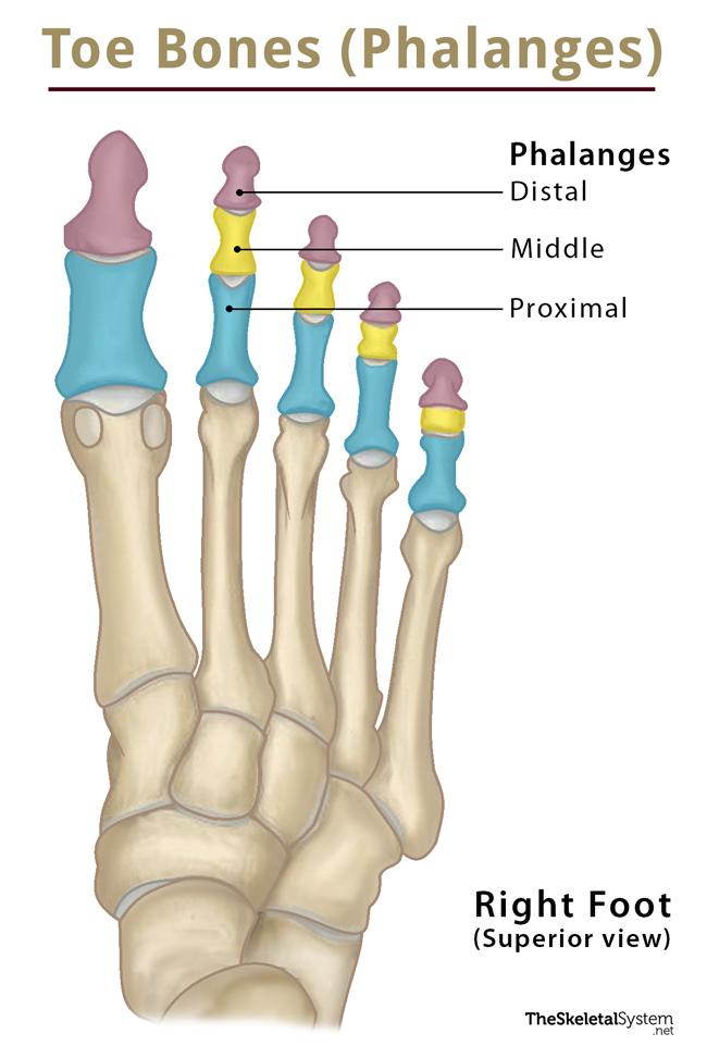 www.theskeletalsystem.net
First Distal Phalanx (toe) Fracture | Image | Radiopaedia.org
 radiopaedia.org
Distal Finger Joint Anatomy Photograph By Maurizio De - Vrogue.co
 www.vrogue.co
[DIAGRAM] Proximal Epiphysis Long Bone Diagram - MYDIAGRAM.ONLINE
![[DIAGRAM] Proximal Epiphysis Long Bone Diagram - MYDIAGRAM.ONLINE](https://www.researchgate.net/publication/330944846/figure/fig2/AS:723873473515528@1549596293938/Bone-macrostructure-a-Growing-long-bone-showing-epiphyses-epiphyseal-plates.png) mydiagram.online
mydiagram.online
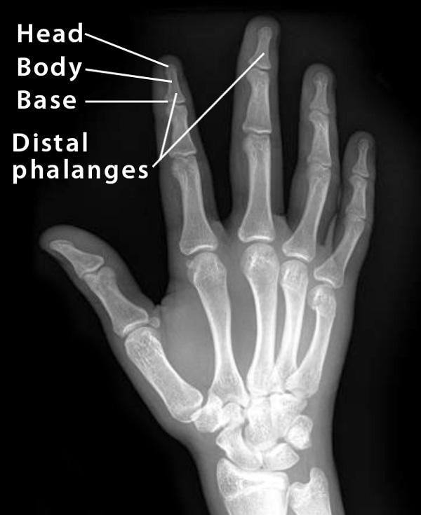
Distal Phalanx: Definition, Location, Anatomy, Diagram
 www.theskeletalsystem.net
distal proximal phalanges hand ray phalanx anatomy there definition ossification diagram location
Ossification Of Bones Of The Hand - Prohealthsys
 www.prohealthsys.com
hand ossification bones anatomy pectoral girdle limb upper figure trapezoid child prohealthsys metacarpals trapezium scaphoid central quizlet
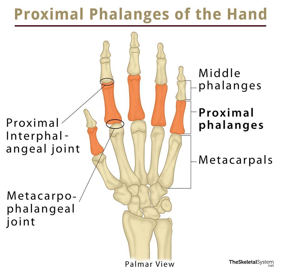
Proximal Phalanx: Definition, Location, Anatomy, Diagram
 www.theskeletalsystem.net
phalanx proximal skeletal anatomy bones location definition system diagram
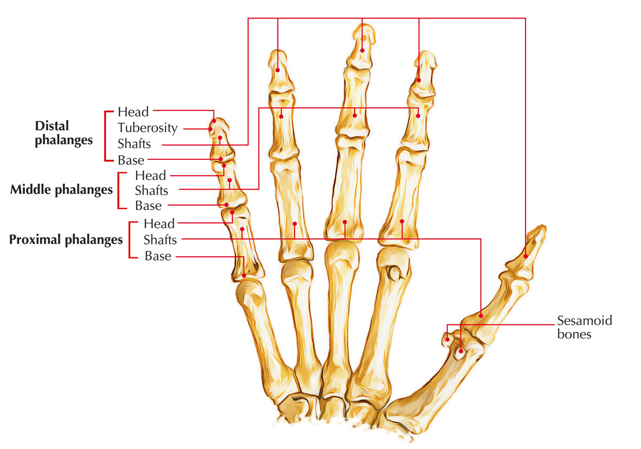
Where Are The Bones Called Phalanges Located
 studybeglerbegs.z4.web.core.windows.net
Skeleton Of The Foot – Earth's Lab
 www.earthslab.com
metatarsal bone bones navicular fifth foot skeleton proximal head phalanges base distal first parts body phalangeal lateral medial fourth end
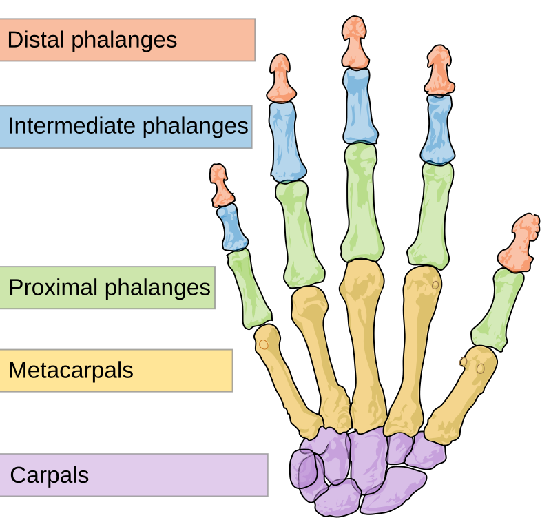
Distal Phalanx - Wikidata
 www.wikidata.org
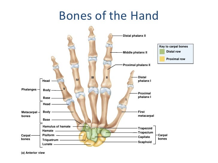
Appendicular Skeleton
 www.slideshare.net
phalanx skeleton appendicular distal bones phalanges metacarpal wrist fracture second capitate anterior injury
X-ray Showing An Avulsion Fracture Of The Proximal Phalanx Of The Thumb
 www.alamy.com
fracture phalanx proximal avulsion fractures articular disruption
Embryology, Anatomy, And Normal Findings | Radiology Key
 radiologykey.com
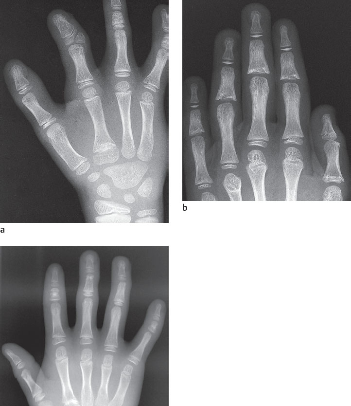
shaped phalanges proximal findings anatomy embryology normal centers ossification toes radiology year old symmetric epiphyseal arrows conical bell third second
16 Malformations And Deformities | Radiology Key
 radiologykey.com
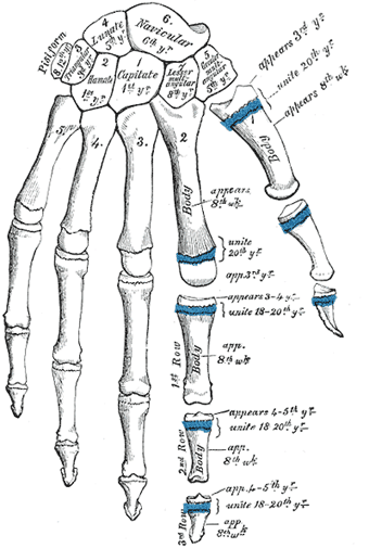
The Phalanges Of The Hand - Human Anatomy
 theodora.com
phalanges hand anatomy bone finger fingers ossification phalanx human third first proximal
Distal Phalanx Fracture
 www.slideshare.net
distal phalanx fracture
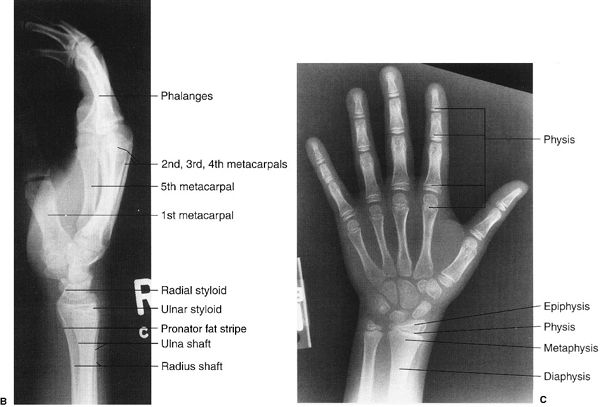
Musculoskeletal System | Radiology Key
 radiologykey.com
hand epiphysis metaphysis physis diaphysis radiograph normal pa lateral right left musculoskeletal system
First distal phalanx (toe) fracture. Epiphysis cleft proximal phalanx radiopaedia radiology. [diagram] proximal epiphysis long bone diagram