← active vs. inactive osteoblasts Osteocytes osteoblasts difference between bone vs cells figure osteoblast cell structure Osteogenic cells and osteoblasts →
If you are looking for Histology at SIU you've visit to the right place. We have 35 Pictures about Histology at SIU like Compact Bone (Decalcified) Series, Osteoblasts_40x, Histo – bone and also Osteoblastoma : Bone Tumor Cancer : Tumors of the bone. Here it is:
Histology At SIU
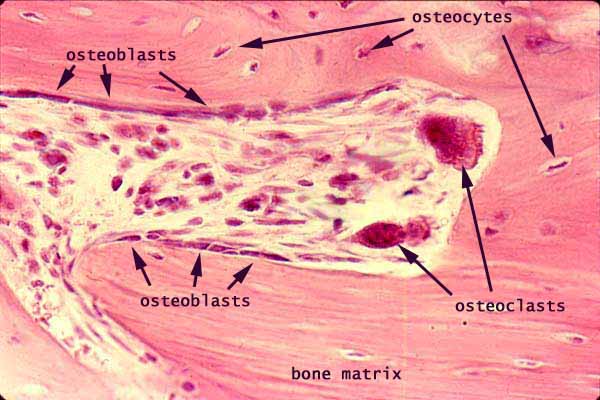 histology.siu.edu
Bone Cells-osteoblasts, Osteocytes And Osteoclasts; Unpublished Image
 www.researchgate.net
osteoblasts osteoclasts osteocytes unpublished filipovic
Osteoblast Histology | Www.pixshark.com - Images Galleries With A Bite!
 pixshark.com
osteoblast histology osteocytes
Osteoblasts - 1.
osteoblasts
H&E Stain (A) Osteoblastoma Showing Proliferating Benign-appearing
 www.researchgate.net
Osteoblast - Wikipedia
 en.wikipedia.org
osteoblast bone wikipedia
Histology Of Connective Tissues Lab
 medcell.org
10.08.08: Histology - Cartilage/Mature Bone
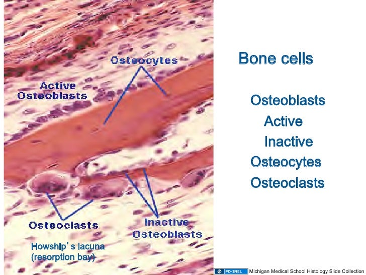 www.slideshare.net
bone histology osteoblasts cartilage inactive osteocytes lacuna osteoclasts cancellous
Histology Of The Bone
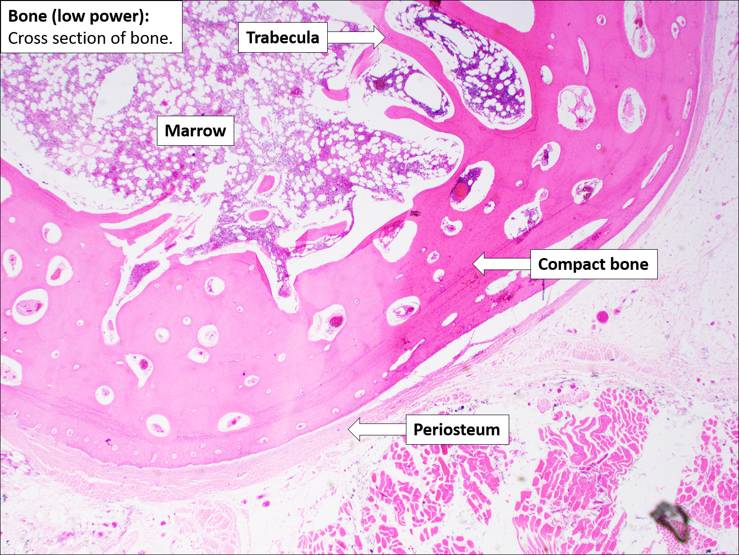 mavink.com
Compact Bone (Decalcified) Series, Osteoblasts_40x
/5_CB_Osteoblasts_40x_a.jpg) www.noelways.com
www.noelways.com
Bone Cells-osteoblasts, Osteocytes And Osteoclasts; Unpublished Image
 www.researchgate.net
osteocytes osteoblasts osteoclasts unpublished filipovic
Defect Formation Group. Activated Osteoblasts Are Aligned Along The
 www.researchgate.net
osteoblasts alveolar defect aligned activated vessels relatively staining observed located
Bone And Bone Formation | Histology
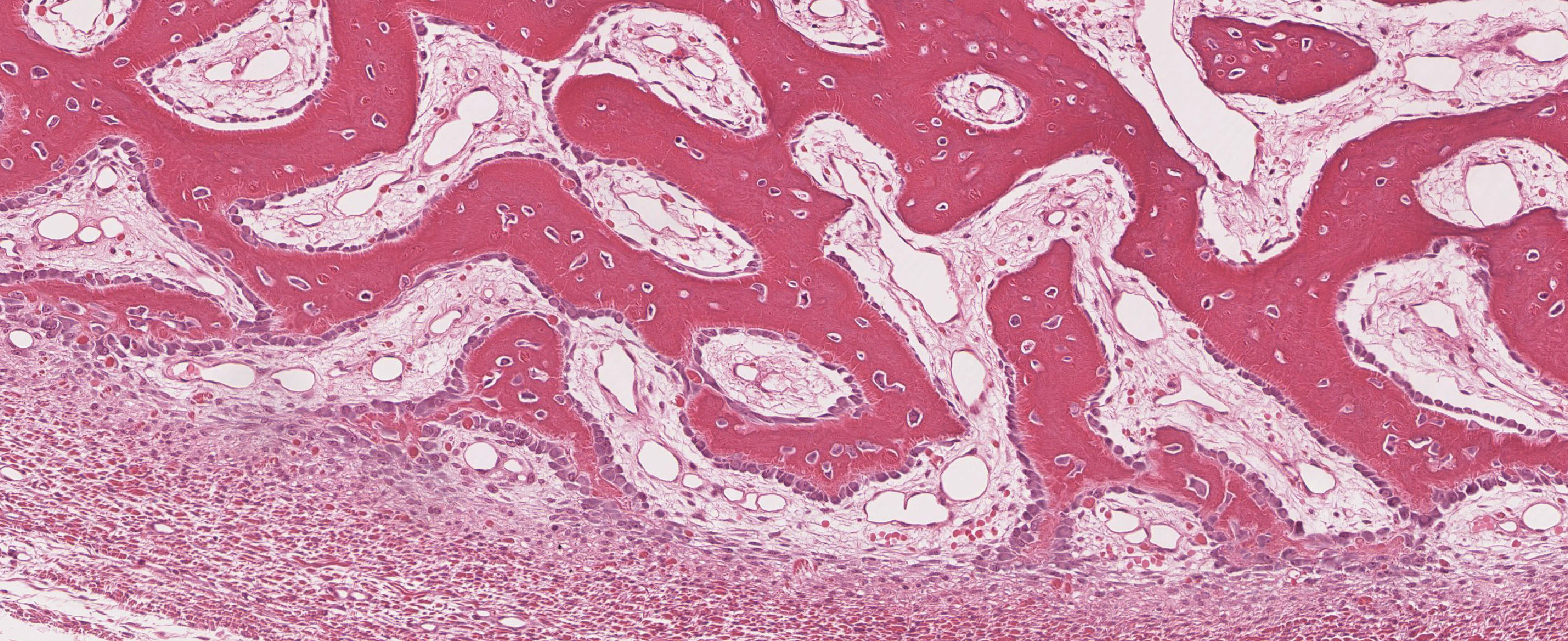 histology.sites.uofmhosting.net
bone histology formation slide slides sites
Osteoblastoma Histology
 ar.inspiredpencil.com
Bone Cells Under A Microscope
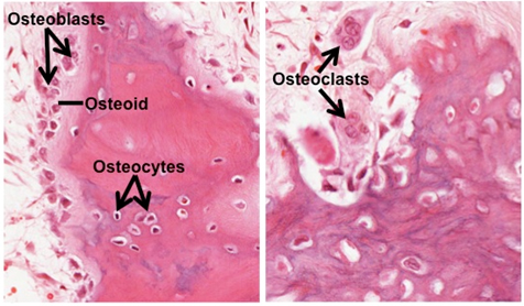 www.animalia-life.club
Histology Of H&E Staining Of The Cartilage Callus In Standard Healing
 www.researchgate.net
Osteoblast | Cell | Britannica
 www.britannica.com
osteoblast osteoblasts developing britannica kids formation pointer magnification
A. Osteoblast B. Osteocytes C. Osteoid D. Cement Line E. Bone | Tissue
 www.pinterest.ph
osteoblast osteoid osteocytes cement bones
Bone Microanatomy – Veterinary Histology
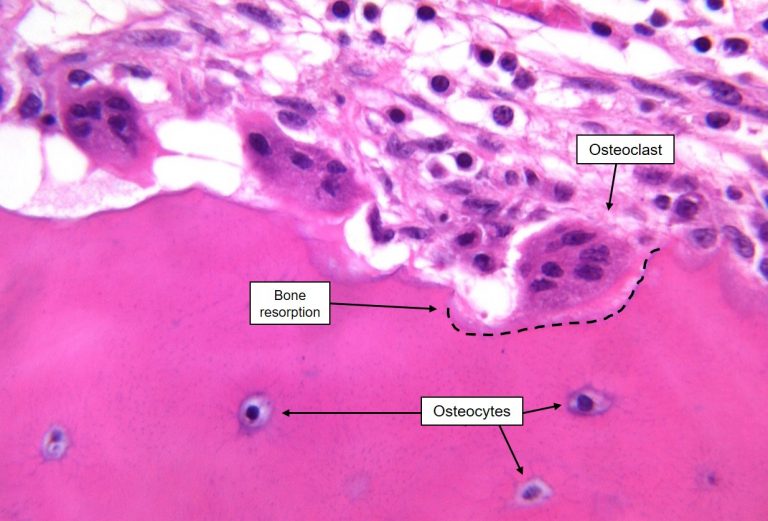 ohiostate.pressbooks.pub
osteoclast microanatomy histology lacuna scalloped pressbooks ohiostate
Endochondral Ossification Labeled
 ar.inspiredpencil.com
00000969 | PEIR Digital Library
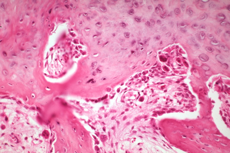 peir.path.uab.edu
00000964 | PEIR Digital Library
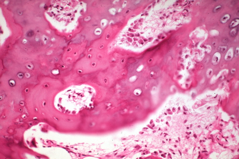 peir.path.uab.edu
Micrograph Showing Some Of The Cellular Components Of Bone (osteocytes
 www.researchgate.net
osteocytes micrograph osteoclast cellular some histology stained
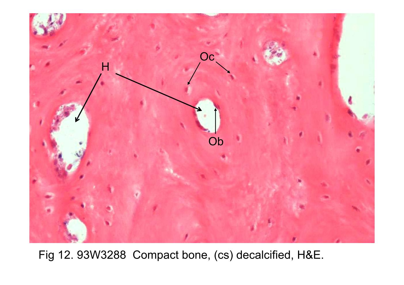
Block6-1/Fig 12. 93W3288 Bone Marrow, Long Bone (cs) H&E.
 anatomy.kmu.edu.tw
bone block6 long decalcified cross cs osteon staining section canal lamellae mature haversian blood osteoblasts marrow fig stained vessels osteocytes
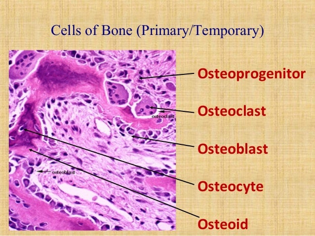
Bone Structure And Clinical Importance
 www.slideshare.net
osteoblast osteoclast osteoprogenitor osteocyte osteoid
-(A) A Typical Histologic Picture Of Osteoblasts In The AG+L-ALD Group
 www.researchgate.net
osteoblasts ald histologic arrows
H&E Histology Showing The Cellular Changes In The Bone Adjacent To The
 www.researchgate.net
Osteoclasts And Osteoblasts Responsible For Bone Resorption And
 www.pinterest.com
osteoblasts osteoclasts anatomy resorption remodeling responsible physiology histology
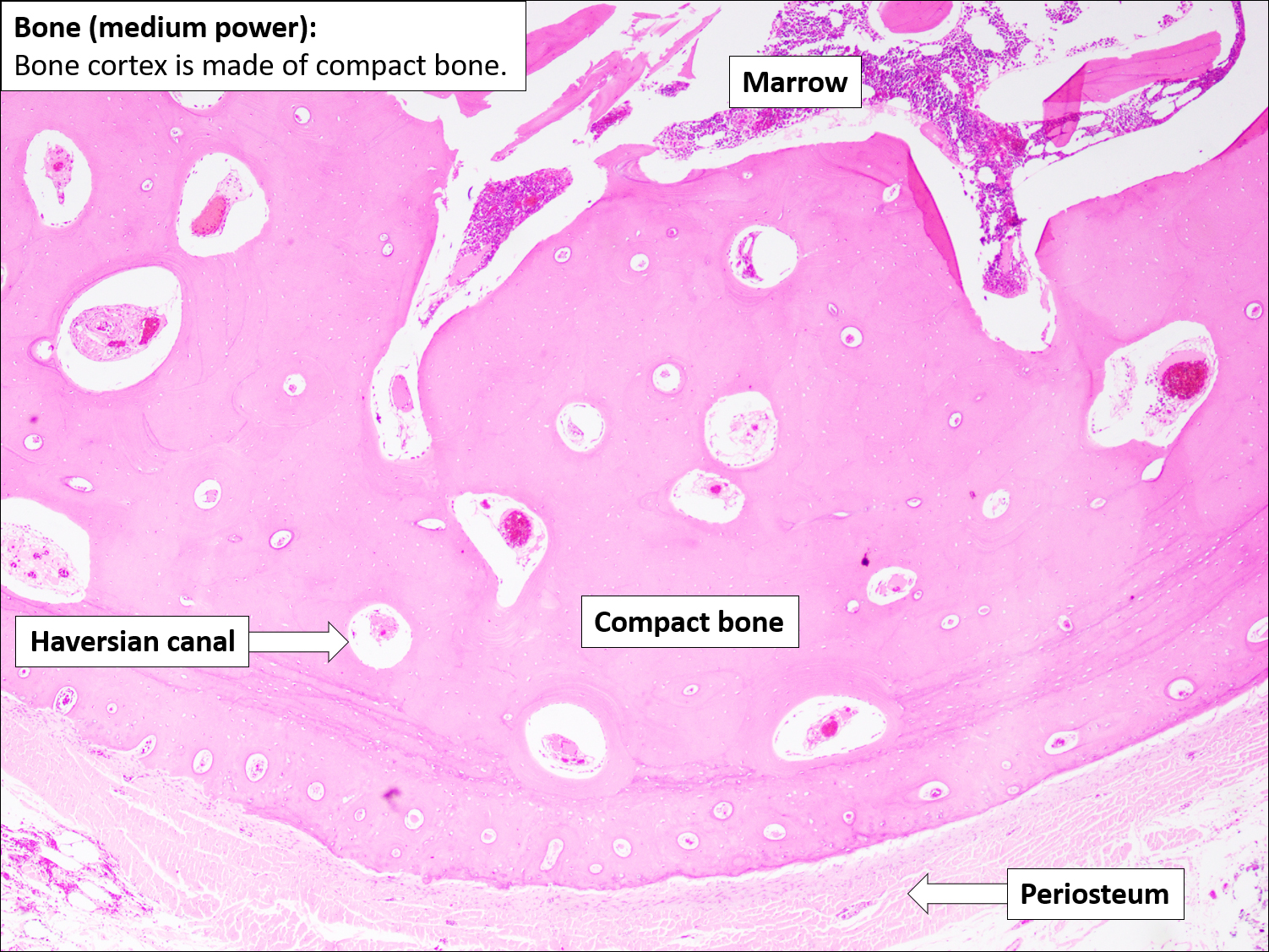
Histology Of Bone Tissue
 mavink.com
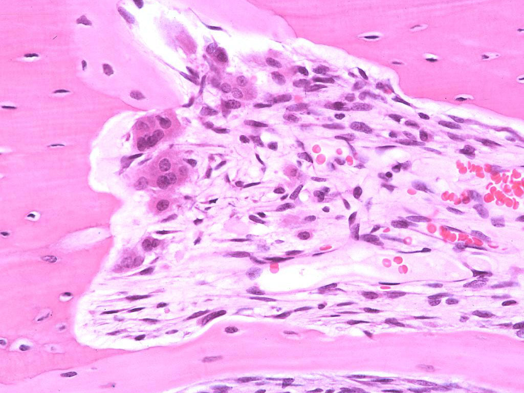
Osteoclasts
 medcell.org
Osteoblastoma : Bone Tumor Cancer : Tumors Of The Bone
 tumorsurgery.org
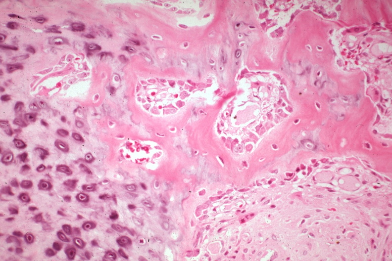
00000971 | PEIR Digital Library
 peir.path.uab.edu
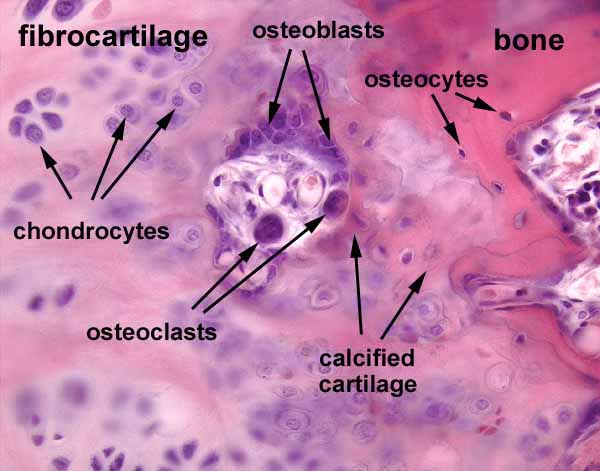
Histology At SIU
 histology.siu.edu
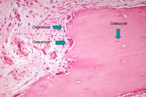
Cells
 elentra.healthsci.queensu.ca
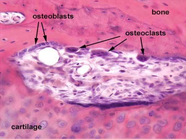
Histo – Bone
 www.slideshare.net
bone histo histology resorption osteoclasts periosteum
Bone histo histology resorption osteoclasts periosteum. H&e stain (a) osteoblastoma showing proliferating benign-appearing. Osteocytes micrograph osteoclast cellular some histology stained