← septum transversum mesenchyme Septum transversum cortical vs trabecular bone Why bone density increases in this area first →
If you are looking for Bone tissue. Coloured scanning electron micrograph (SEM) of human you've came to the right web. We have 35 Pics about Bone tissue. Coloured scanning electron micrograph (SEM) of human like Cortical bone, light micrograph - Stock Image - C052/1948 - Science, Concept of Education anatomy and physiology of Compact bone, or and also Compact Bone | Human anatomy and physiology, Microscopic photography. Here it is:
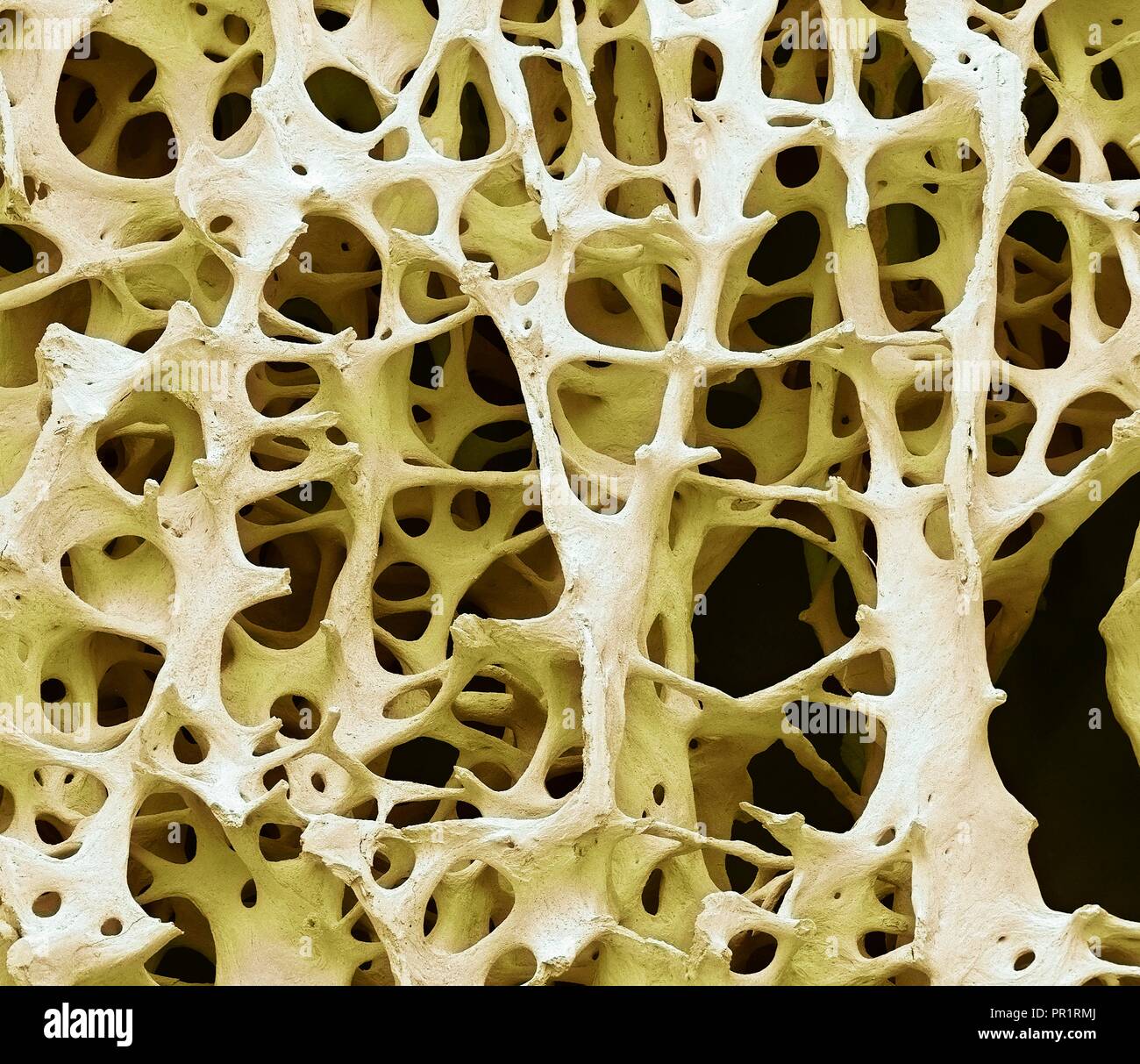
Bone Tissue. Coloured Scanning Electron Micrograph (SEM) Of Human
 www.alamy.com
bone tissue spongy cancellous human sem trabeculae compact cortical magnification shaped rod electron network micrograph marrow spaces substance forming x13
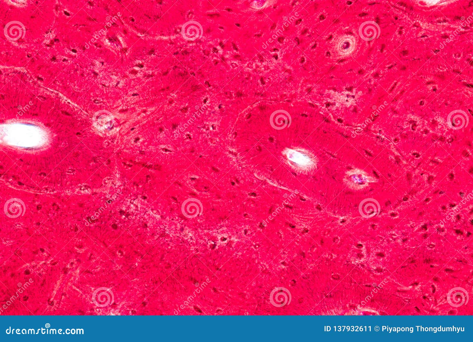
Concept Of Education Anatomy And Physiology Of Compact Bone, Or
 www.dreamstime.com
cortical anatomy physiology
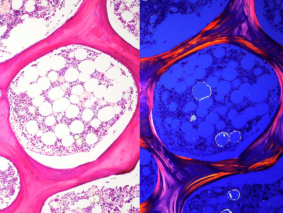
Cancellous Bone Photograph By Jose Calvo / Science Photo Library - Fine
 fineartamerica.com
cancellous calvo jose anatomical
Cortical Bone Histology
 ar.inspiredpencil.com
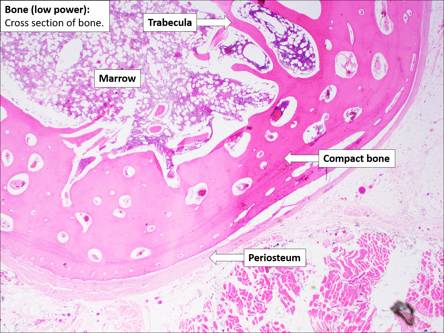
Bone – Normal Histology – NUS Pathweb :: NUS Pathweb
 medicine.nus.edu.sg
histology nus pathweb annotations medicine
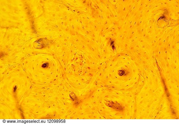
Cortical Bone Tissue Showing Haversian Canals Cortical Bone Tissue
 www.imageselect.eu
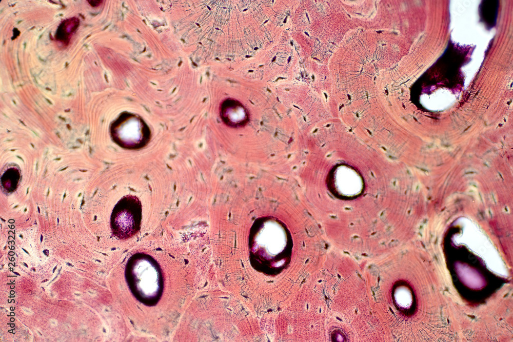
Histology Of Human Compact Bone Tissue Under Microscope View For
 stock.adobe.com
Compact Bone | Human Anatomy And Physiology, Microscopic Photography
 www.pinterest.com
bone microscopic cortical tissue bones osseous histology physiology synonymous
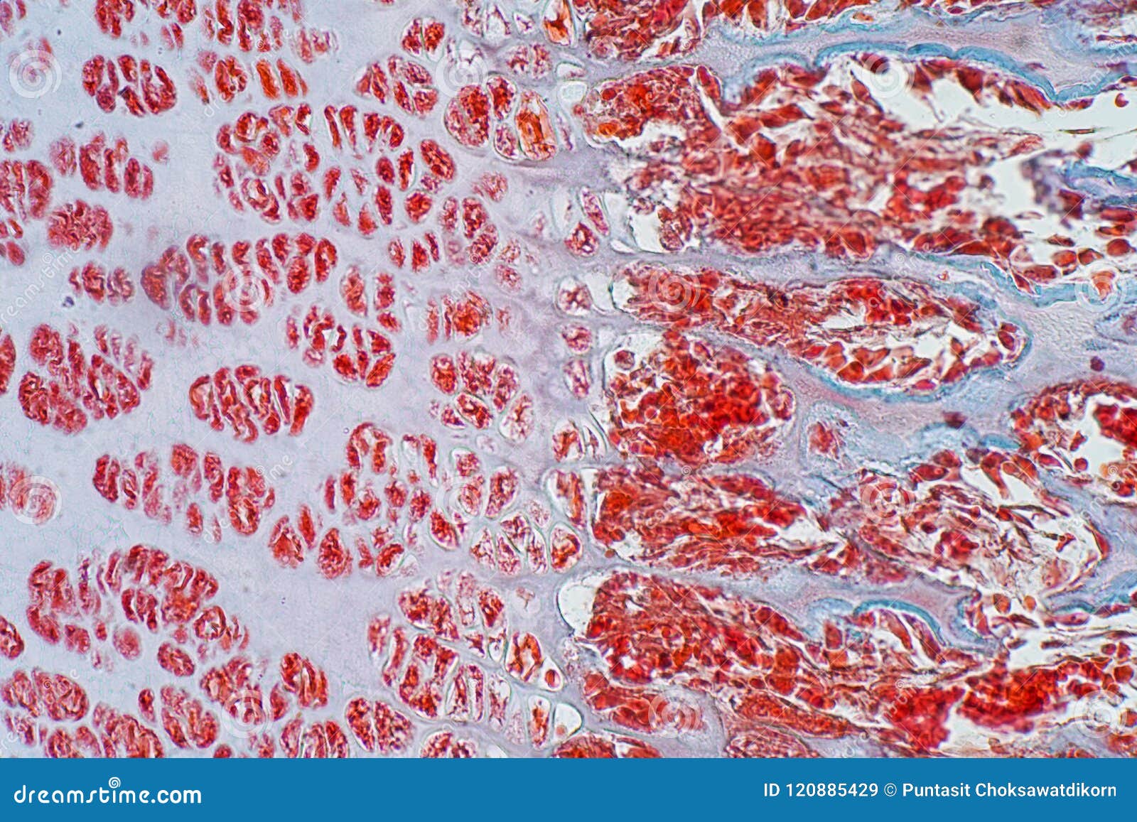
Microscopic Cross Section Of A Bone - Connective Tissue Microscope
 delmav-maniac.blogspot.com
microscope histology maniac structure
CaptionBone Tissue. Coloured Scanning Electron Micrograph (SEM) Of
 www.pinterest.com.mx
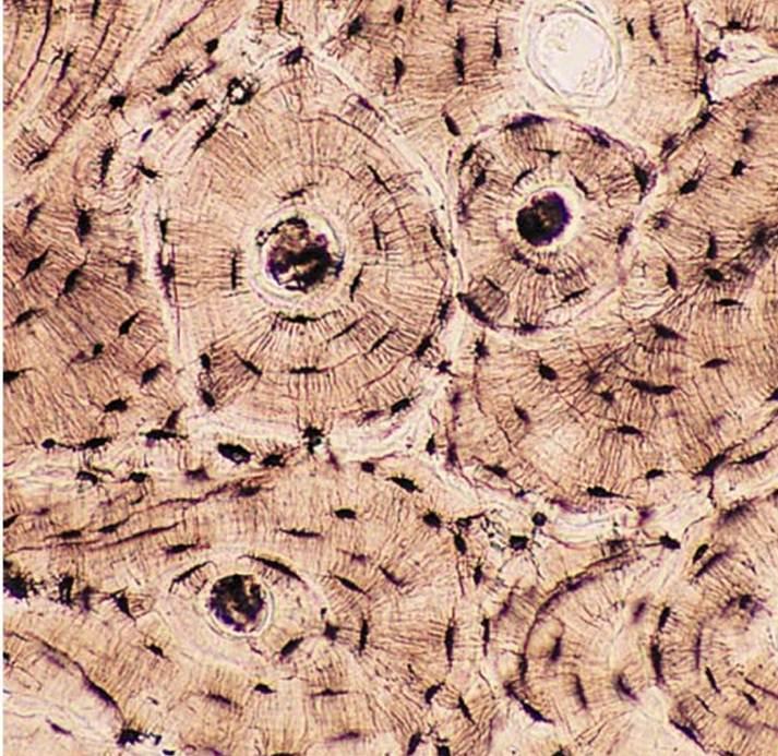
Bone Connective Tissue Under Microscope
 www.animalia-life.club
Bone Cross Section Under Microscope : Backscattered Scanning Electron
 michals-capful.blogspot.com
bone microscope microscopic electron
Bone Cross Section Under Microscope / Cross Section Human Image Photo
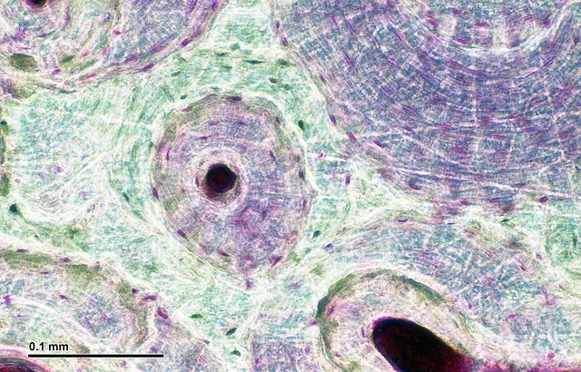 crochetandocomzani.blogspot.com
microscope under tissue section human 400x magnification collagen involves placing trial simply
Bone – Normal Histology – NUS Pathweb :: NUS Pathweb
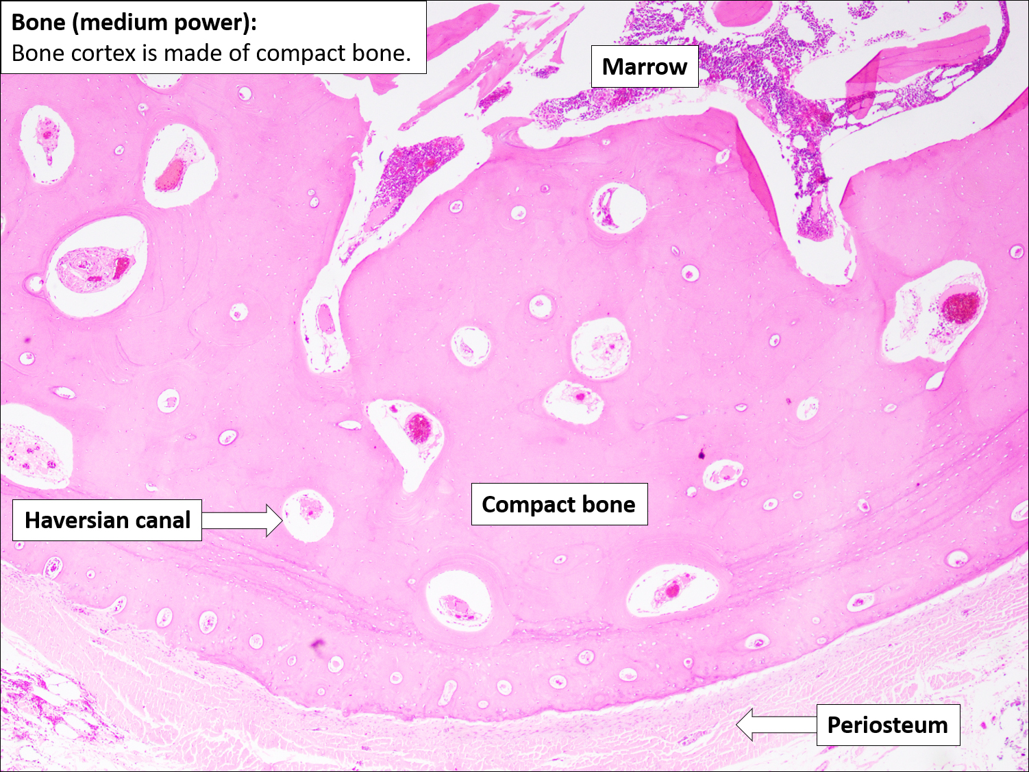 medicine.nus.edu.sg
histology annotations
Bone Tissue And Cells Under The Microscope
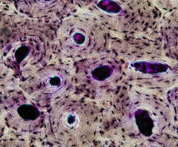 www.microscopemaster.com
bone compact section cross tissue histology microscope under cells cc ground
Chapter 6: CONNECTIVE TISSUE – Human Anatomy (MASTER)
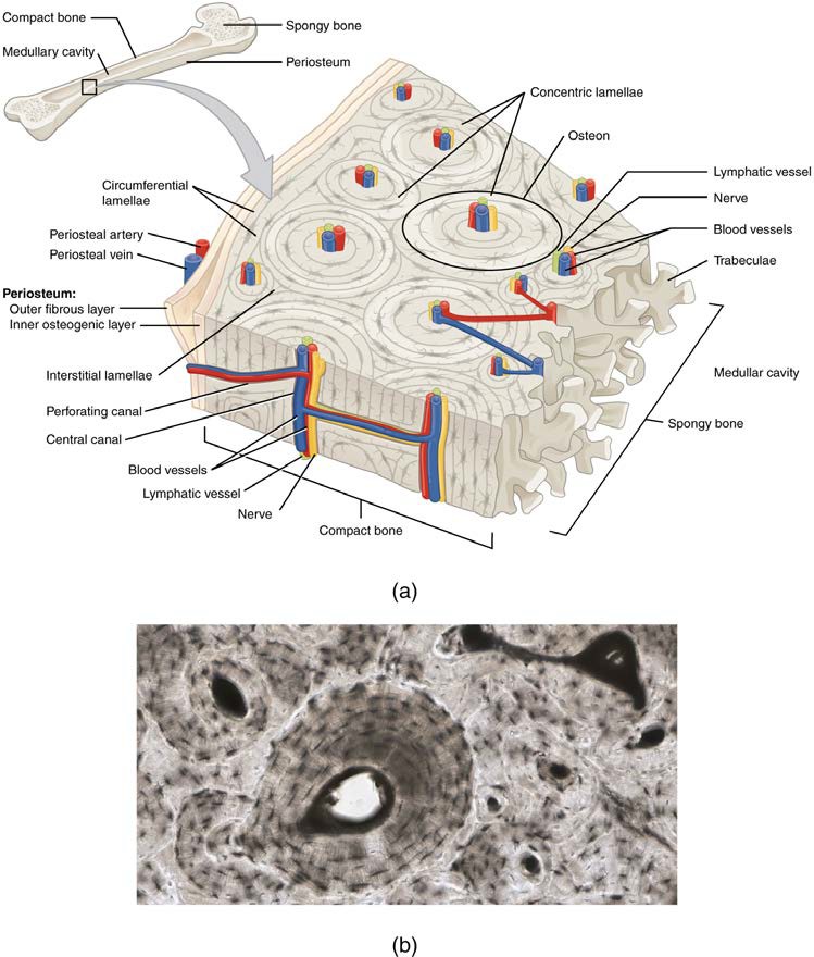 qut.pressbooks.pub
Cortical Bone, Light Micrograph - Stock Image - C052/1948 - Science
 www.sciencephoto.com
Concept Of Education Anatomy And Physiology Of Compact Bone, Or
 www.dreamstime.com
Micrograph Spongy Bone Hi-res Stock Photography And Images - Alamy
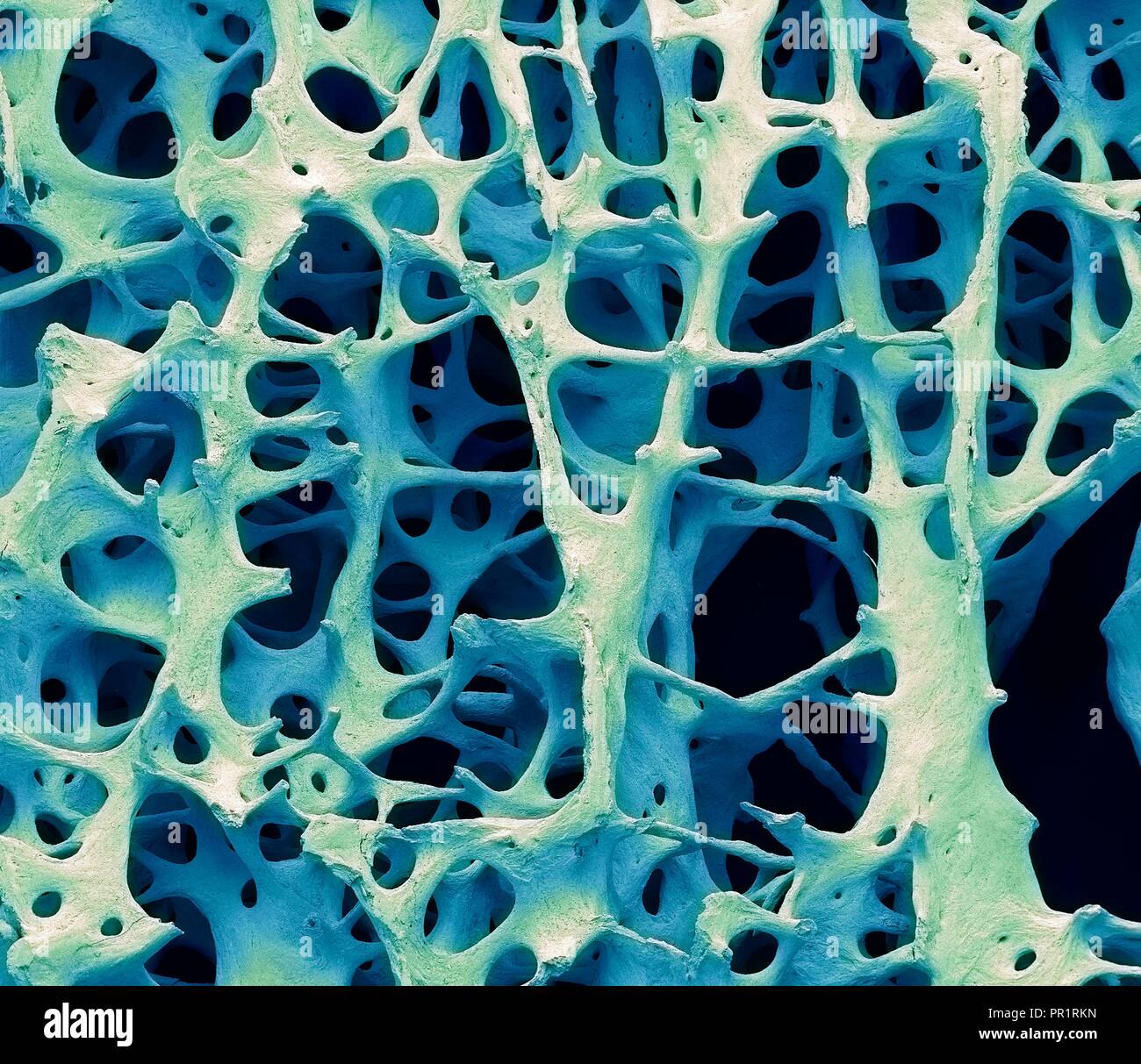 www.alamy.com
Human Kidney, Cortical Zone Under Microscope Stock Photo - Alamy
 www.alamy.com
kidney microscope under cortical
A-C Ultrastructure Of Bone. A Light Microscope Image Of A Ground
 www.researchgate.net
Print Anatomy Tissue Practical Review Flashcards | Easy Notecards
 www.easynotecards.com
photomicrograph histology skeletal slides cortical bones x100 allposters
Cortical Bone Tissue Royalty-Free Stock Photo | CartoonDealer.com
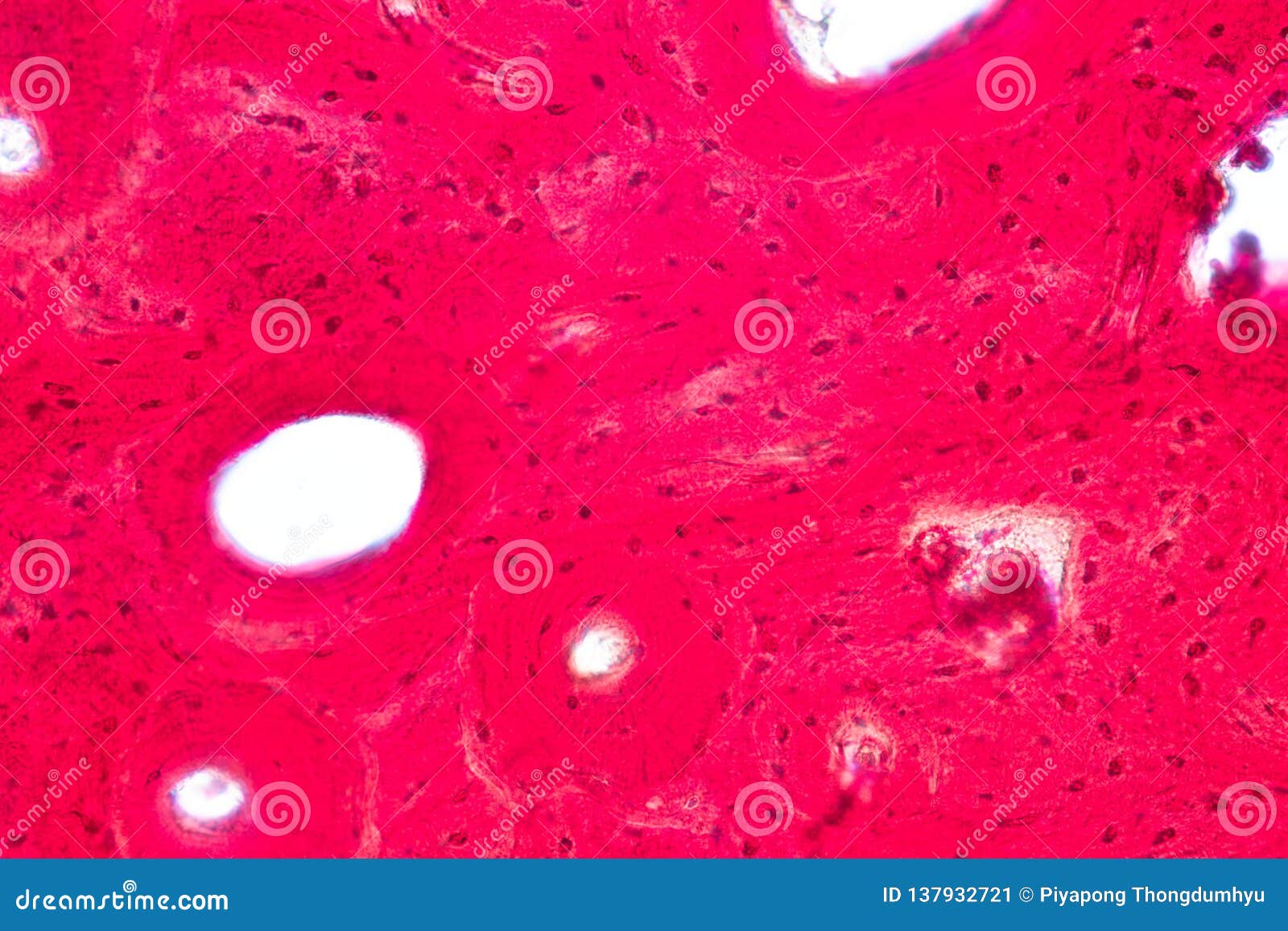 cartoondealer.com
Concept Of Education Anatomy And Physiology Of Compact Bone, Or
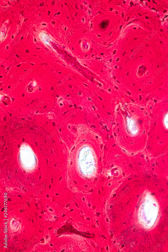 stock.adobe.com
Cortical Bone Tissue Showing Haversian Canals, Osteons, Osteocytes And
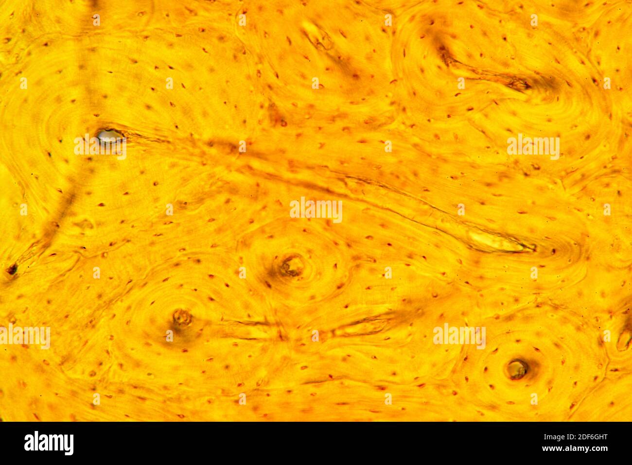 www.alamy.com
Compact Bone Microscope Labeled
 anatomybrainley57.netlify.app
6.3C: Microscopic Anatomy Of Bone - Medicine LibreTexts
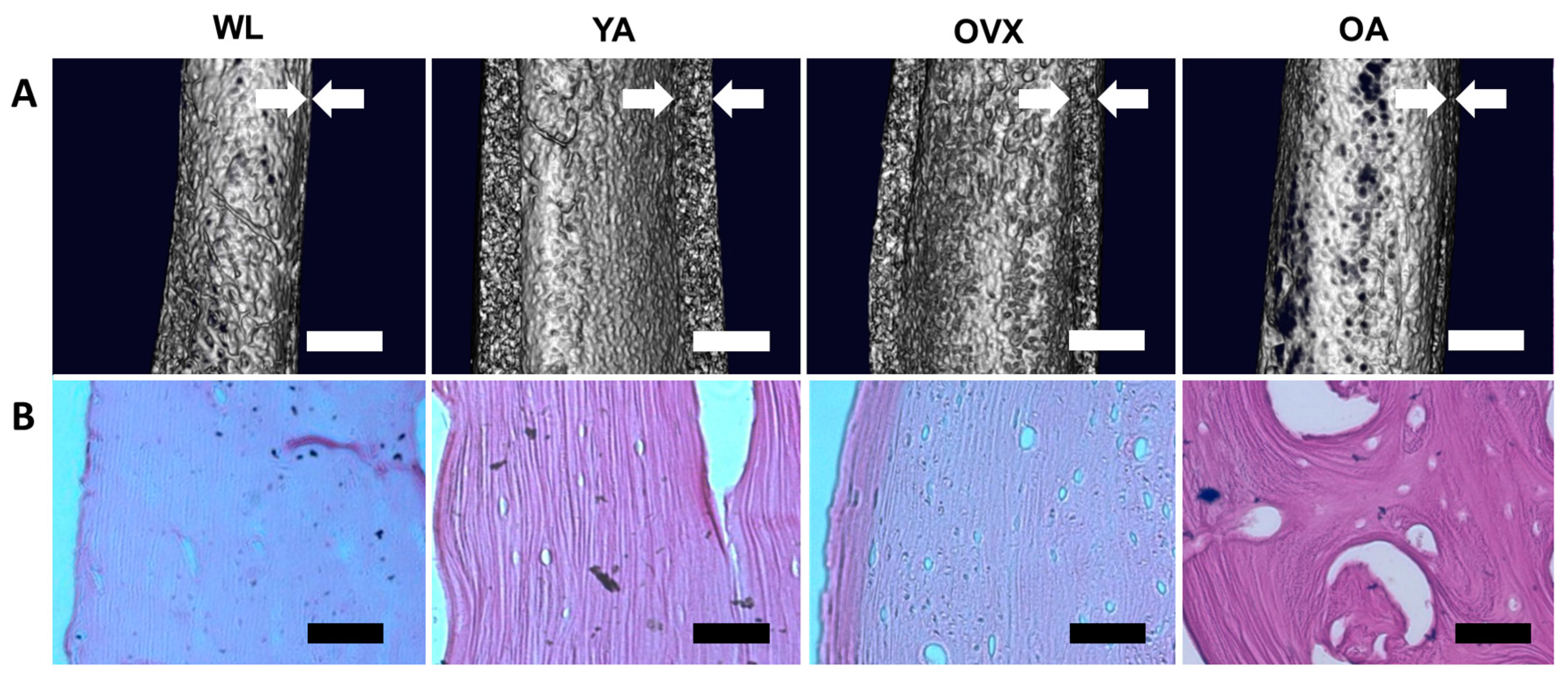
Bioengineering | Free Full-Text | Assessment Of The Inner Surface
 www.mdpi.com
cortical microstructure bioengineering assessment decellularized microscope scanning electron
Photomicrograph Of Compact Bone Labeled
 mavink.com
65 Cortical Bone Images, Stock Photos & Vectors | Shutterstock
 www.shutterstock.com
BONE HISTOLOGY
 microanatomy.net
bone compact histology microanatomy canal haversian osteocytes lacunae osteon rib entire layers lined several unit above shows their
Bone Cross Section Under Microscope - / The Cortical Area Is A Measure
 curlyjessie.blogspot.com
bone microlabgallery microscope
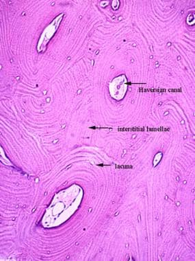
Histology Of Bone: Background, Gross Structure Of Long Bone, Nerves And
 emedicine.medscape.com
Cortical Bone Photograph By Jose Calvo / Science Photo Library - Fine
 fineartamerica.com
Bone Connective Tissue Under Microscope
 www.animalia-life.club
Bone cross section under microscope / cross section human image photo. Micrograph spongy bone hi-res stock photography and images. Kidney microscope under cortical