← cortical compact bone Concept of education anatomy and physiology of compact bone, or diaphysis of long bone Bone cronin jerry dr file diaphysis long →
If you are searching about Case 264: A Case of Osseous Sarcoidosis | Radiology you've came to the right page. We have 35 Pics about Case 264: A Case of Osseous Sarcoidosis | Radiology like anteroposterior radiographs of the left wrist, showing slight cortical, (A) CT bone setting images showing cortical thinning and irregularity and also Adamantinoma | Radiology Reference Article | Radiopaedia.org. Read more:
Case 264: A Case Of Osseous Sarcoidosis | Radiology
 pubs.rsna.org
sarcoidosis osseous radiology radiol
Osteoporosis. Multilevel Lumbar Spine Cortical Thinning And Lack Of
 www.researchgate.net
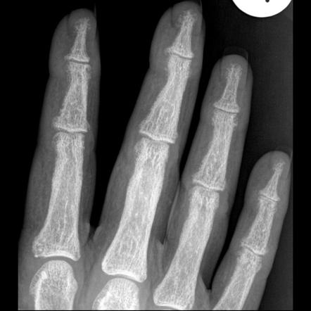
Subperiosteal Bone Resorption | Radiology Reference Article
 radiopaedia.org
resorption subperiosteal hyperparathyroidism radiology tuft radiopaedia erosions erosion distal parathyroid thyroid
Roentgen Ray Reader: Cortical Tunneling
 roentgenrayreader.blogspot.com
cortical tunneling bone ray striations cortex
Bone Structure Cortical (Compact) Cancellous (trabecular, Spongy) - Ppt
+Cancellous+(trabecular%2C+spongy).jpg) slideplayer.com
slideplayer.com
bone cortical cancellous trabecular compact spongy structure tissue long trabeculae between composition density plate surface subchondral rods porosity marrow plates
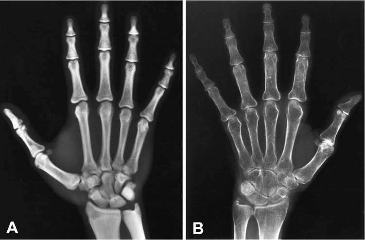
Severe Osteoporosis X Ray
 animalia-life.club
A CT Scan Of The Knee Reveals A Cortical Bone Defect. | Download
 www.researchgate.net
Magnification Of A Small, Incomplete Subtrochanteric Fracture With
 www.researchgate.net
fracture incomplete cortical femur subtrochanteric magnification thickening demonstrates
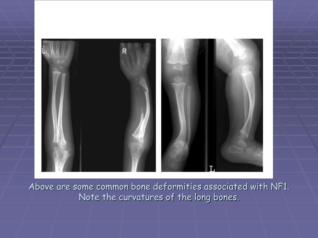
PPT - Neurofibromatosis Type 1 PowerPoint Presentation, Free Download
 www.slideserve.com
nf1 neurofibromatosis bone deformities bones long curvatures note associated common above some ppt type powerpoint presentation
The Characteristic X Ray Findings: Slender Long Bones With Thick Cortex
 www.researchgate.net
bones cortex slender findings characteristic thick dysplasia fibrous tibia medullary
Plain Radiographs Of Both Hands Showing Cortical Thickening Of II, III
 www.researchgate.net
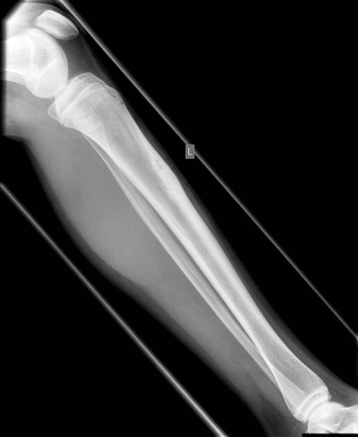
Adamantinoma | Radiology Reference Article | Radiopaedia.org
 radiopaedia.org
Surgical Neurology International
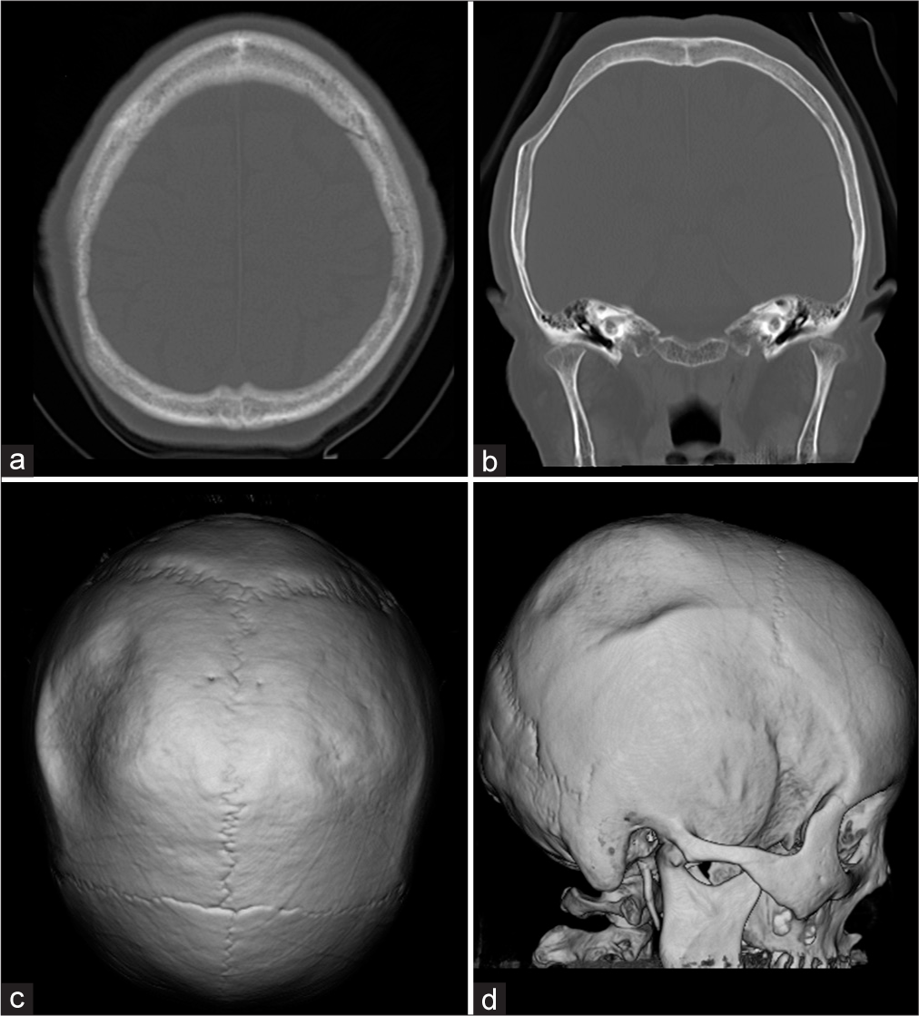 surgicalneurologyint.com
Cross-section Of Human Femur Showing Trabecular And Cortical Bone From
 www.researchgate.net
cortical trabecular femur section human
Micro-CT Imaging Examples Illustrating A Cortical Bone Thinning Of The
 www.researchgate.net
Neurofibromatosis - Mind Map
 www.mindomo.com
Marked Cortical Thickening Of The Radius And Ulna. | Download
 www.researchgate.net
cortical thickening ulna radius marked
Thinning Bones
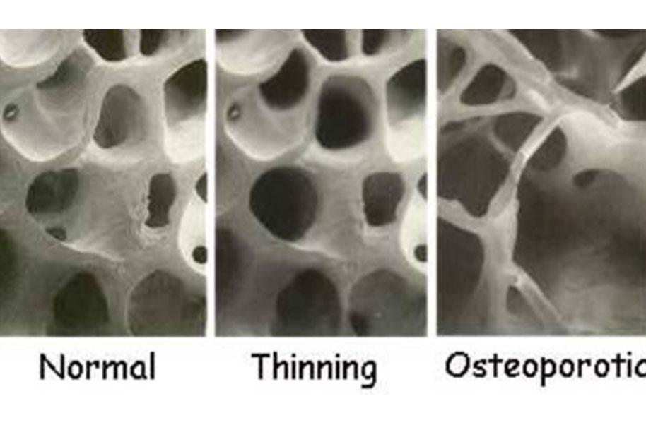 www.yogavanahill.com
bones thinning bone
B: Bone With Cortical Thinning, Bone Demineralization And Multiple
 www.researchgate.net
X-ray Showed A Tumor Of The Right Ischium With Thinning Of The Cortical
 www.researchgate.net
Cortical Lesions Of The Tibia: Characteristic Appearances At
 pubs.rsna.org
cortical tibia lesions characteristic conventional appearances radiography old proximal
How To Identify Normal Nucleus & Annulus In MRI Of IVD? – Digital Teaching
 digitalteaching.org
cortical bone compact tissue cortex gif mri normal dense less
Irresti: Cortical Desmoid Knee Mri
 irresistable89.blogspot.com
cortical desmoid mri humerus radiographic correlation irresti
PPT - Lecture # 13: Bone Tissue PowerPoint Presentation, Free Download
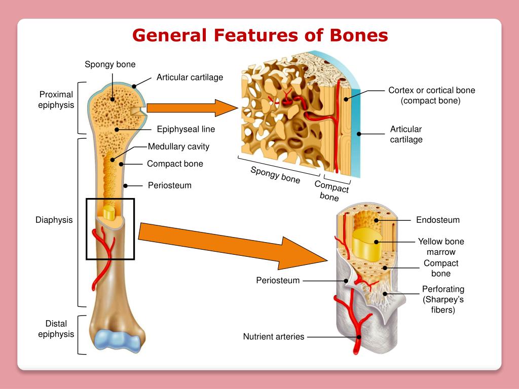 www.slideserve.com
bone endosteum tissue periosteum medullary spongy cavity perforating marrow cartilage
(A) CT Bone Setting Images Showing Cortical Thinning And Irregularity
 www.researchgate.net
Association Of Cortical Shape Of The Mandible On Panoramic Radiographs
 www.semanticscholar.org
mandible cortical bone figure trabecular beam cone association shape mandibular radiographs panoramic ct adults structure analysis japanese
Surgical Neurology International
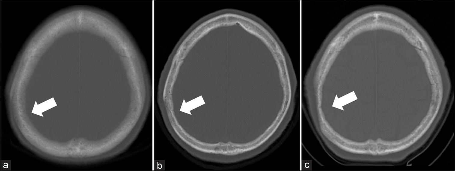 surgicalneurologyint.com
Radiographs Of Long Bone Showing Cortical Thickening With Medullary
 www.researchgate.net
21: Metabolic Diseases, Nutritional Disorders, And Skeletal Dysplasias
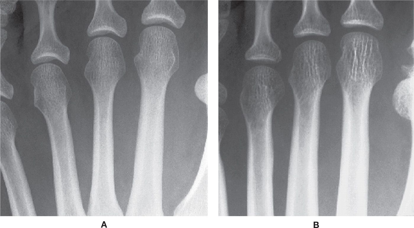 musculoskeletalkey.com
skeletal diseases dysplasias nutritional metabolic disorders osteopenia cortical thinning bone normal coarse osteoporosis endosteal chronic resorption figure musculoskeletalkey
(A) CT Bone Setting Images Showing Cortical Thinning And Irregularity
 www.researchgate.net
Generalized Rarefaction Of Jaw Bones /prosthodontic Courses
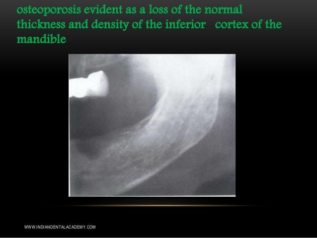 www.slideshare.net
jaw generalized rarefaction bones prosthodontic osteoporosis
13.1: Introduction To Bone Health - Medicine LibreTexts
 med.libretexts.org
spongy libretexts
Cortical Versus Trabecular Bone - My Endo Consult
 myendoconsult.com
Cortical Bone Loss Is An Early Feature Of Nonradiographic Axial
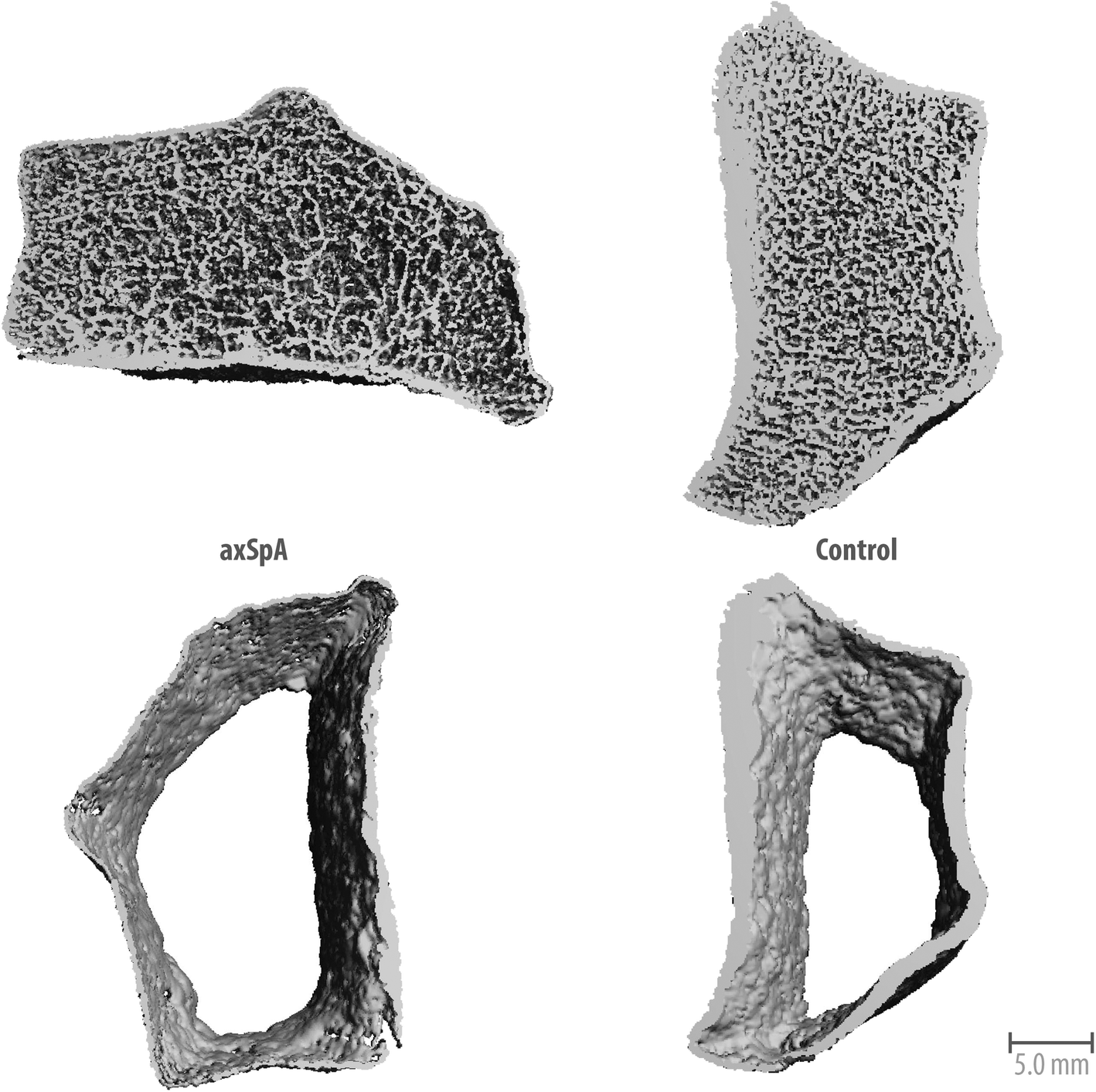 arthritis-research.biomedcentral.com
bone cortical loss spondyloarthritis axial feature fig early 1620
Anteroposterior Radiographs Of The Left Wrist, Showing Slight Cortical
 www.researchgate.net
Fracture incomplete cortical femur subtrochanteric magnification thickening demonstrates. (a) ct bone setting images showing cortical thinning and irregularity. Case 264: a case of osseous sarcoidosis