← hyaline cartilage hip-joint Hip joint anatomy • easy explained crayon de papier dessin Crayon joli vont aider archzine améliorer →
If you are searching about Ultrasound images of hyaline-vascular variant CD. a Longitudinal image you've came to the right place. We have 35 Pictures about Ultrasound images of hyaline-vascular variant CD. a Longitudinal image like ___Gouty Arthritis # Calcification along the superior layer of hyaline, Musculoskeletal Ultrasound | Radiology Key and also Figure 3 from Knee ultrasound in pediatric patients – anatomy. Read more:
Ultrasound Images Of Hyaline-vascular Variant CD. A Longitudinal Image
 www.researchgate.net
Ultrasound Images Before And After Cavitation In An MCP Joint. From
 www.researchgate.net
___Gouty Arthritis # Calcification Along The Superior Layer Of Hyaline
 www.pinterest.com.mx
Hyaline Cartilage, Light Micrograph - Stock Image - P174/0036 - Science
 www.sciencephoto.com
hyaline cartilage micrograph light
Hyaline Cartilage Involvement In Patients With Gout And Calcium
 www.oarsijournal.com
calcium disease ultrasound deposition pyrophosphate
1, Chondro-osseous Junction Between The Bony Part And The Cartilaginous
 www.pinterest.com
ultrasound cartilage hyaline part cartilaginous bony femoral junction chondro osseous between choose board
Figure 3 From Knee Ultrasound In Pediatric Patients – Anatomy
 www.semanticscholar.org
An Ultrasound Image Of Femoral Hyaline Cartilage, A: Normal, B: Mild
 www.researchgate.net
Musculoskeletal - MSK Ultrasound - Insight Medical Imaging
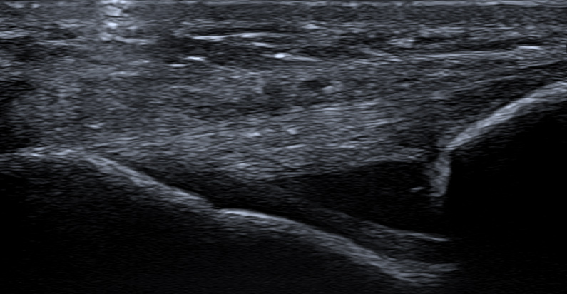 x-ray.ca
ultrasound msk musculoskeletal medical
| Healthy Hyaline Cartilage. Dorsal Longitudinal (A) And Transverse (B
 www.researchgate.net
Ultrasound Case Of The Month Nov 13
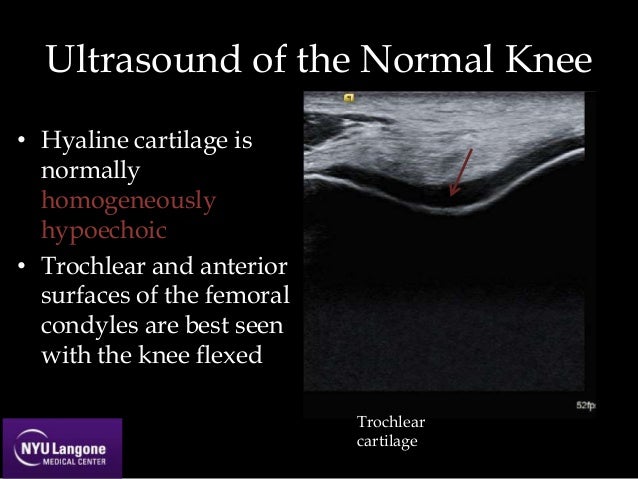 www.slideshare.net
ultrasound cartilage hyaline trochlear
Hyaline Cartilage, Light Micrograph - Stock Image - C032/0446 - Science
 www.sciencephoto.com
cartilage hyaline micrograph light
Musculoskeletal Ultrasound | Radiology Key
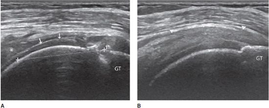 radiologykey.com
ultrasound musculoskeletal tendon figure
A. Ultrasound GS Short Axis Image Of The Femoral Hyaline Cartilage
 www.researchgate.net
femoral cartilage hyaline axis ultrasound
-Enchondroma. Lobules Of Hyaline Cartilage With Thin Mantle Of Fibrous
 www.researchgate.net
Musculoskeletal Ultrasound | Radiology Key
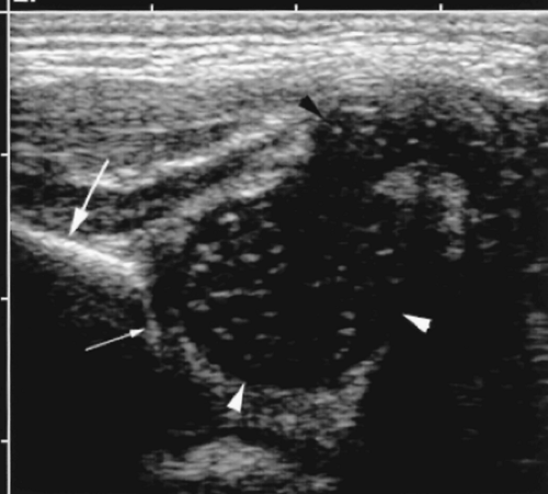 radiologykey.com
ultrasound cartilage musculoskeletal hyaline
A. Ultrasound GS Short Axis Image Of The Femoral Hyaline Cartilage
femoral cartilage hyaline gs ultrasound fully t2 weighed flexed
Hyaline Cartilage
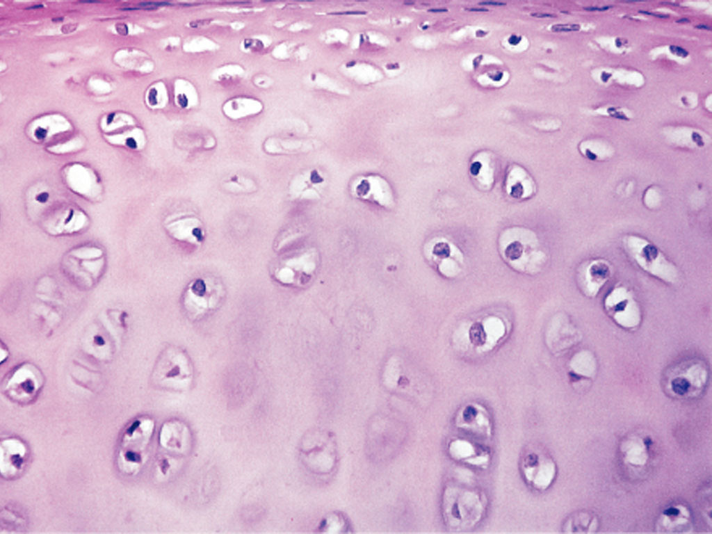 medcell.org
Mammal. Hyaline Cartilage. Transverse Section. 250X - Hyaline Cartilage
 www.nature-microscope-photo-video.com
August 2013: NYU MSK Ultrasound Case Of The Month
 www.slideshare.net
ultrasound msk costochondral junction cartilage nyu
Pin By Anette Mnabhi On MSK US | Ultrasound, Sonography, Medical
 www.pinterest.com
cartilage hyaline
Cartilage – Meyers Histology
 histology-online.com
Gout - Internet Book Of MSK Ultrasound
 mskultrasound.net
Hyaline Cartilage, Light Micrograph - Stock Image - C047/7320 - Science
 www.sciencephoto.com
LM Of A Section Through Hyaline Cartilage - Stock Image - P174/0027
 www.sciencephoto.com
cartilage hyaline section histology lm tissue anatomy through lab low sciencephoto identify
Ultrasound Image Of ISLN Block. (A) Hyoid Bone And Thyroid Cartilage
 www.researchgate.net
MSK Ultrasound Shoulder Orange County - Huntington Beach Sports
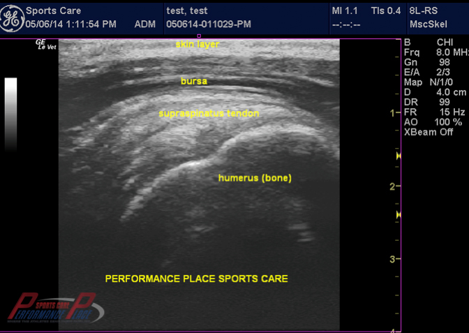 www.p2sportscare.com
ultrasound shoulder msk read normal anatomy supraspinatus scapula tear cuff labeled rotator knee facts pain radiology
Isogenous Groups In Hyaline Cartilage, Light Micrograph - Stock Image
 www.sciencephoto.com
Deep Learning-Based Femoral Cartilage Automatic Segmentation In
 www.umbjournal.org
Ultrasound Case Of The Month Nov 13
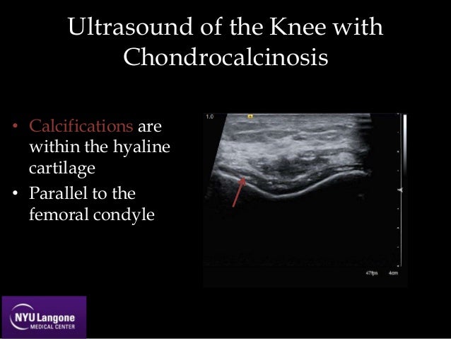 www.slideshare.net
ultrasound chondrocalcinosis femoral
Imaging Of Other Tissues | Radiology Key
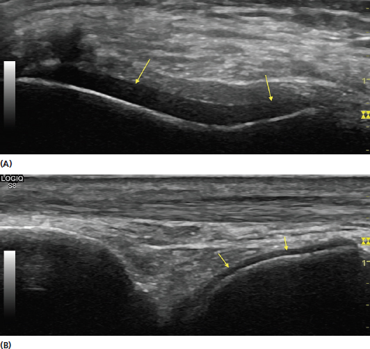 radiologykey.com
tissues imaging other cartilage demonstrating bone sonograms joint hypoechoic hyaline figure radiologykey
Musculoskeletal Ultrasound | Radiology Key
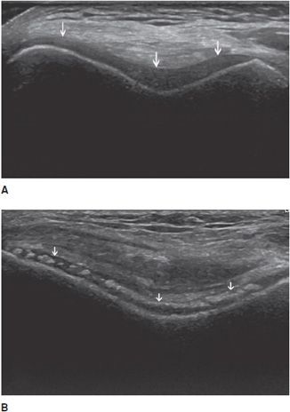 radiologykey.com
ultrasound musculoskeletal trochlear cartilage hyperechoic hyaline hypoechoic normal figure
Healthy Subjects. Pictorial Evidence Of Ultrasound Normal And Abnormal
 www.researchgate.net
MSK Ultrasonography | Diagnostic Ultrasound | Whitstable Kent
 www.joylaneclinic.co.uk
ultrasound knee msk ultrasonography pain sound muscle tendon use ligaments shoulder joint inside body tissue soft ray like
Human Hyaline Cartilage, Sec. 7 µm H&E Microscope Slide | Carolina
 www.carolina.com
cartilage hyaline microscope slide human sec carolina µm slides histology
-enchondroma. lobules of hyaline cartilage with thin mantle of fibrous. Cartilage hyaline micrograph light. Calcium disease ultrasound deposition pyrophosphate