← periosteum human body diagram Periosteum outer and inner layer osteocytes in spongy bone Spongy bone photos and premium high res pictures →
If you are searching about Intramembranous Ossification Stock Image - Image of histology you've visit to the right page. We have 35 Pictures about Intramembranous Ossification Stock Image - Image of histology like Periosteum and bone, light micrograph - Stock Image - C026/4045, Histology, Periosteum And Endosteum | Treatment & Management | Point of and also Periosteum Definition, Location, Anatomy, Histology and Function. Read more:
Intramembranous Ossification Stock Image - Image Of Histology
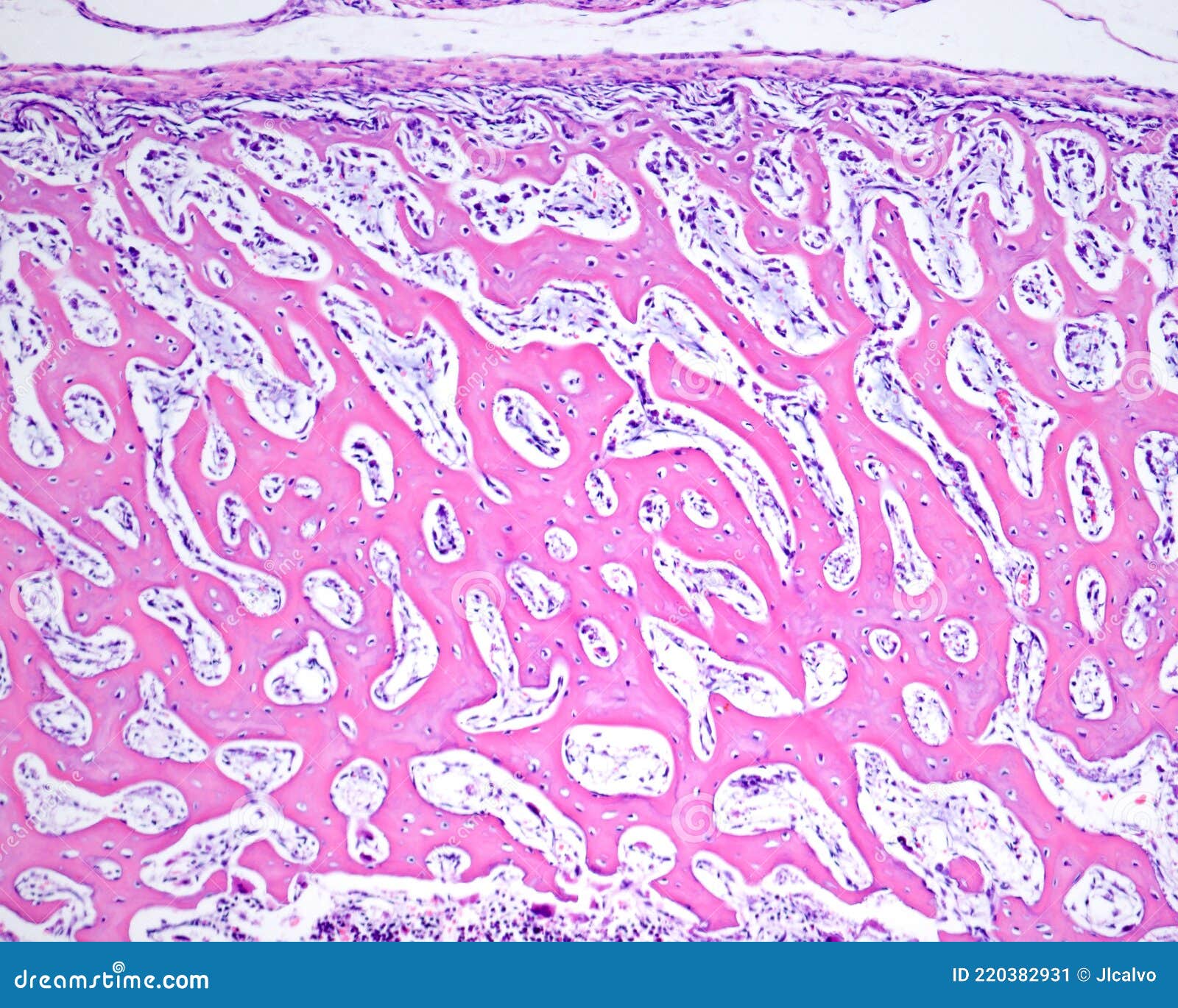 www.dreamstime.com
Endosteum : Definition, Function, Histology, Vs Periosteum - (updated
 healthfixit.com
endosteum histology periosteum section tissue cavity vessel medullary connective
Bone Histology - Embryology
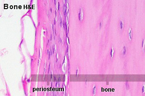 embryology.med.unsw.edu.au
bone periosteum histology endosteum embryology development au musculoskeletal
Traumatized Periosteum: Its Histology, Viability, And Clinical
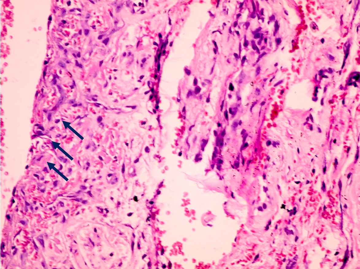 orthopedicreviews.openmedicalpublishing.org
Compared With Adult Periosteum, Infant Periosteum, As Depicted In This
(PDF) The Periosteum. Part 1: Anatomy, Histology And Molecular Biology
 www.researchgate.net
periosteum histology periosteal midshaft abundance femoral arrowheads comprising cambium surface masson μm stained trichrome
Periosteum Stok Fotoğraf, Resimler Ve Görseller - IStock
 www.istockphoto.com
Normal Periosteum Diagram | Quizlet
 quizlet.com
Histology, Periosteum And Endosteum | Treatment & Management | Point Of
 www.statpearls.com
Periosteum Stock Photos, Pictures & Royalty-Free Images - IStock
 www.istockphoto.com
compacto histologia periosteum bone tissue histology osso humano microscope ósseo connective sob microscópio
10.08.08: Histology - Cartilage/Mature Bone
 www.slideshare.net
histology periosteum endosteum cartilage histo
[PDF] The Periosteum. Part 1: Anatomy, Histology And Molecular Biology
![[PDF] The periosteum. Part 1: Anatomy, histology and molecular biology](https://d3i71xaburhd42.cloudfront.net/1f4dc57f426e15961d4df579c3f4a17eebc7461c/5-Figure2-1.png) www.semanticscholar.org
www.semanticscholar.org
[조직학 이야기] 뼈 : 네이버 블로그
![[조직학 이야기] 뼈 : 네이버 블로그](http://postfiles15.naver.net/20150319_270/yeol0513_1426752019677WL06r_JPEG/bc_fig_10_e.jpg?type=w2) blog.naver.com
blog.naver.com
Periosteum Stock Photos, Pictures & Royalty-Free Images - IStock
 www.istockphoto.com
periosteum stock microscope under bone videos
Pacinian Corpuscle In Periosteum #1 Photograph By Jose Calvo / Science
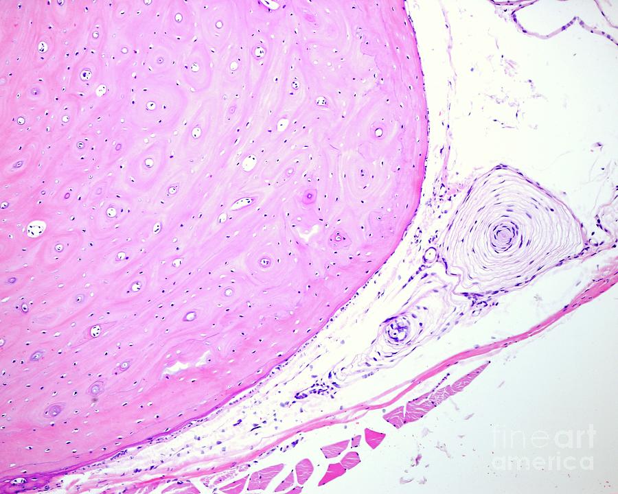 pixels.com
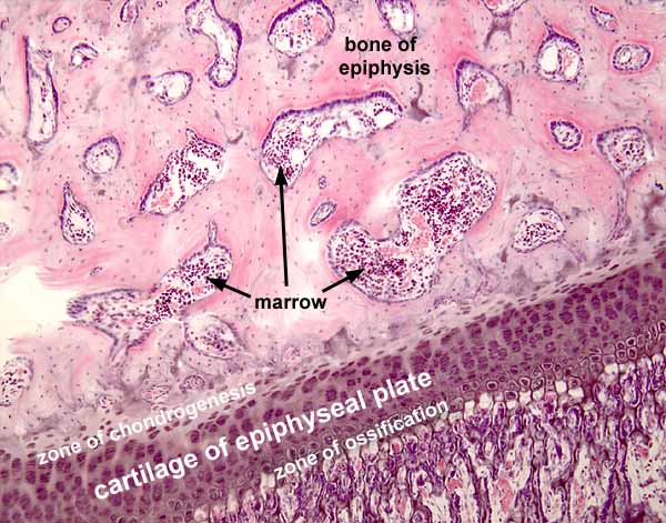
Histology At SIU
 histology.siu.edu
Periosteum Histology
 ar.inspiredpencil.com
Periosteum Histology Slide Diagram | Quizlet
 quizlet.com
SPONGY BONE HISTOLOGY | Microanatomy Web Atlas | Gwen V. Childs, Ph.D.
 microanatomy.net
bone histology spongy periosteum microanatomy cannon where layers osteogenic osteoblasts cells htm
(a) Conventional Light Microscopy. Hematoxylin And Eosin-stained
 www.researchgate.net
Microscopic Bone Anatomy Diagram
 schematiclibmammees88.z22.web.core.windows.net
Dimensional Change Of The Healed Periosteum On Surgically Created Defects
 www.jpis.org
periosteum figure dimensional surgically healed defects created change jpis
Periosteum Stock Photos, Pictures & Royalty-Free Images - IStock
 www.istockphoto.com
Histology, Periosteum And Endosteum | Treatment & Management | Point Of
 www.statpearls.com
SPONGY BONE HISTOLOGY | Microanatomy Web Atlas | Gwen V. Childs, Ph.D.
 microanatomy.net
bone spongy compact histology osteon microanatomy tissue marrow slide rib space web spaces osteoblasts cartilage found part atlas question lacuna
Figure 1 From The Periosteum. Part 1: Anatomy, Histology And Molecular
 www.semanticscholar.org
periosteum histology
Periosteum And Bone, Light Micrograph - Stock Image - C026/4045
 www.sciencephoto.com
periosteum micrograph
160+ Periosteum Stock Photos, Pictures & Royalty-Free Images - IStock
 www.istockphoto.com
Periosteum Photos Stock Photos, Pictures & Royalty-Free Images - IStock
 www.istockphoto.com
Periosteum Definition, Location, Anatomy, Histology And Function
 www.ehealthstar.com
periosteum anatomy tissue connective diagram ligaments tendons location dense histology ehealthstar irregular bone fibers function medical definition sharpey picture spaces
Histology, Periosteum And Endosteum - StatPearls - NCBI Bookshelf
 www.ncbi.nlm.nih.gov
periosteum endosteum ncbi histology compact
Strength And Histological Characteristics Of Periosteal Fixation To
 www.liebertpub.com
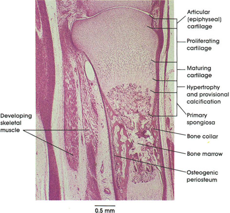
Endochondral Bone Formation Histology
 animalia-life.club
Photomicrograph Of Compact Bone Labeled
 mavink.com
Histological Analysis Of Periosteum (P) And IM Samples. (A-F) Analysis
 www.researchgate.net
periosteum histological fibrous layer bv
Periosteum stok fotoğraf, resimler ve görseller. Periosteum and bone, light micrograph. Figure 1 from the periosteum. part 1: anatomy, histology and molecular