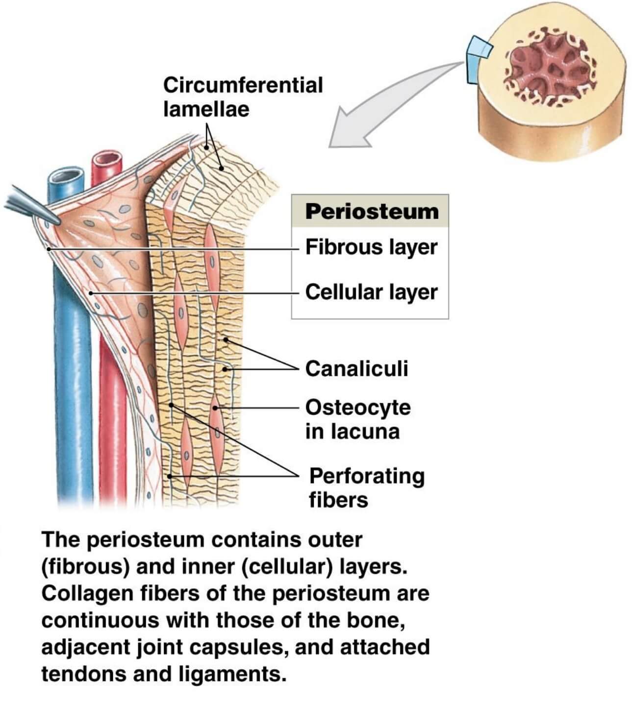
← periosteum fibrous layer Periosteum function anatomy periosteum light microscope Stevenel's blue/ van gieson's picrofuchsin stained samples for light →
If you are searching about 9- Oral mucosa (1) – Composition and layers - Dr Wadhah Oral histology you've came to the right web. We have 35 Images about 9- Oral mucosa (1) – Composition and layers - Dr Wadhah Oral histology like Layers of Oral Mucosa Diagram | Quizlet, Oral mucosa and also Inspect Oral Cavity Patterns With Narrow Band Imaging (NBI) - Olympus. Read more:
9- Oral Mucosa (1) – Composition And Layers - Dr Wadhah Oral Histology
 www.youtube.com
mucosa oral histology layers composition
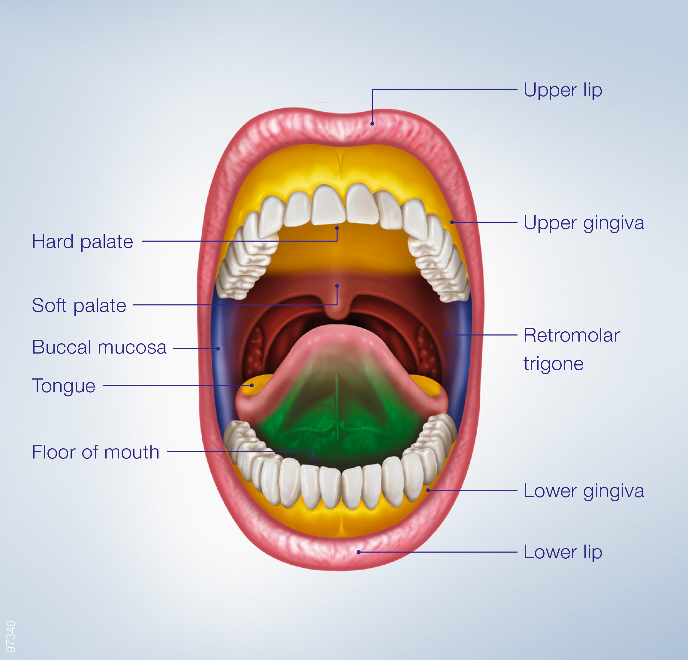
Inspect Oral Cavity Patterns With Narrow Band Imaging (NBI) - Olympus
 www.olympusprofed.com
oral cavity epithelium inspect
Where Is Epithelium Connective Tissue And Blood Vesse - Vrogue.co
 www.vrogue.co
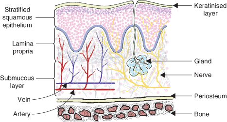
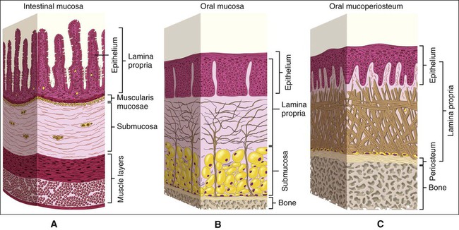
1 Oral Embryology, Histology And Anatomy | Pocket Dentistry
 pocketdentistry.com
oral mucosa histology diagram structure anatomy kind embryology tissues permission reproduced ireland figure pocketdentistry
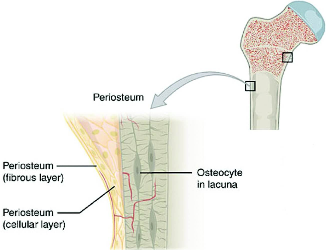
Periosteum Function Anatomy
 studybeglerbegs.z4.web.core.windows.net
[Figure, Oral Mucosa, Epithelium, Lamina Propria,...] - StatPearls
![[Figure, Oral Mucosa, epithelium, lamina propria,...] - StatPearls](https://www.ncbi.nlm.nih.gov/books/NBK565867/bin/Oral_Mucosa2_Outlines-01.jpg) www.ncbi.nlm.nih.gov
www.ncbi.nlm.nih.gov
Structure Of Oral Mucous Membrane | Oral Mucous Membrane |Oral Mucosa #
 www.youtube.com
Ppt - Oral Mucosa Powerpoint Presentation - Id:482082 F42
 mungfali.com
Figure 1 From The Periosteum. Part 1: Anatomy, Histology And Molecular
 www.semanticscholar.org
periosteum histology
Oral Audie – Telegraph
 telegra.ph
Layers Of Oral Mucosa Diagram | Quizlet
 quizlet.com
Когда возникает воспаление надкостницы зуба: причины и лечение флюса
 dentazone.ru
Figure 1 From Periosteum: A Highly Underrated Tool In Dentistry
 www.semanticscholar.org
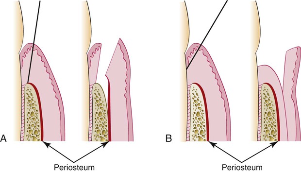
57: The Periodontal Flap | Pocket Dentistry
 pocketdentistry.com
flap periodontal thickness full incision partial bevel flaps internal mucosal diagram dentistry figure reflection bone reflect pocket note after exposure
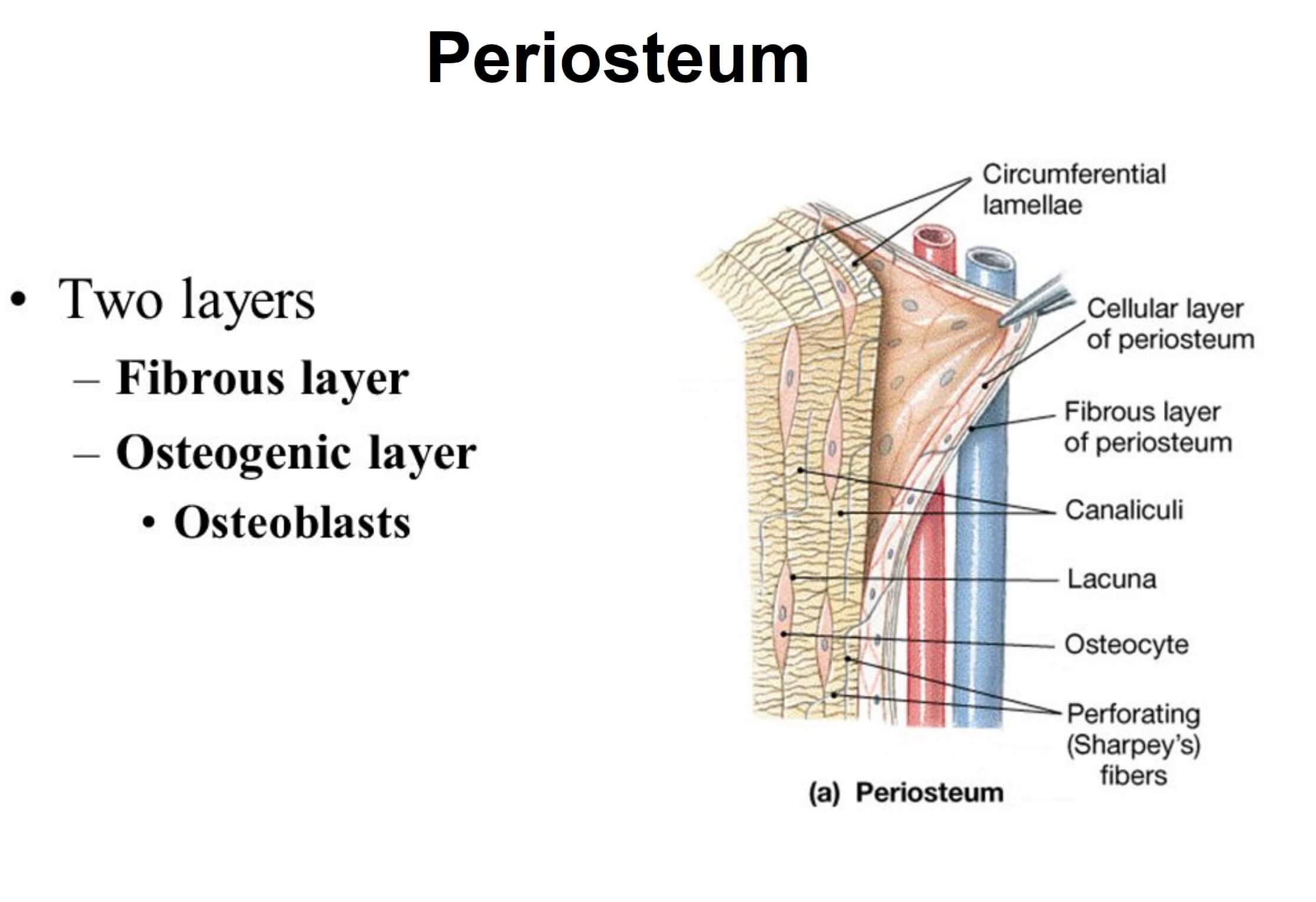
What Is Periosteum? | News | Dentagama
 dentagama.com
periosteum tissue covering dentagama bones dentistry dental connective meaning osteoblasts nerves peri covers
The Periosteum For Regenerating Periodontal Lesions | SteinerBio™
 www.steinerbio.com
periosteum bone histology periodontal lesions regenerating underlying
Normal And Hyperplastic Oral Mucosa Flashcards | Quizlet
 quizlet.com
Shows A Cross Section Of Normal Buccal Mucosa Illustrating The
 www.researchgate.net
mucosa buccal illustrating fig5
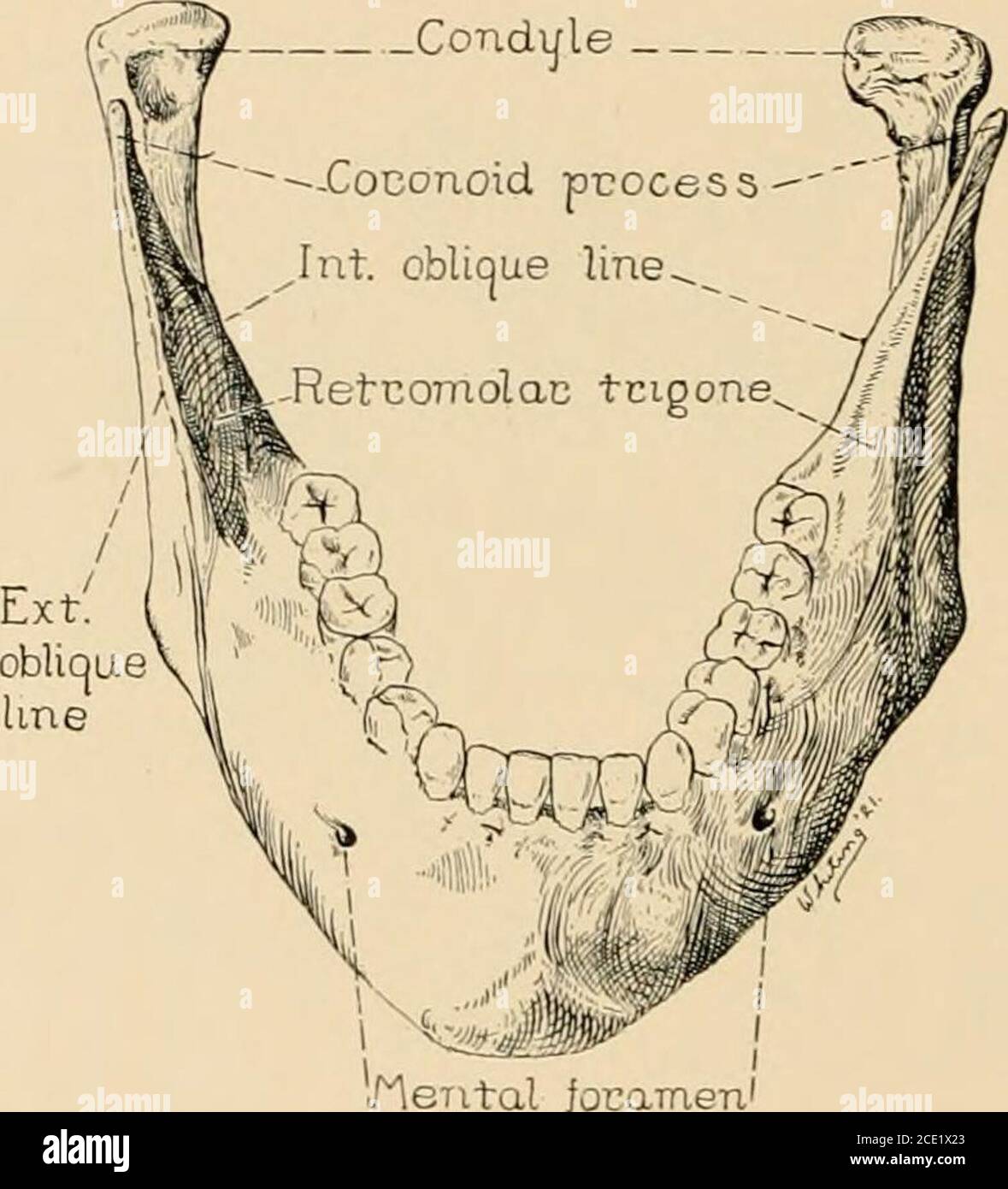
. Regional Anesthesia : Its Technic And Clinical Application . Uscle
 www.alamy.com
Structure And Functions Of The Oral Mucosa SpringerLink, 56% OFF
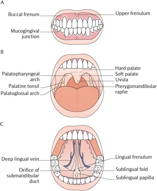 gbu-taganskij.ru
FIG. 1-2 View Of Vestibule. A Change In Color At The Mucogingival
 www.pinterest.ca
dental vestibule labial maxillary mucogingival frenum mandibular cavity pocketdentistry
Clinical Appearance Of Gingiva: A) Attached Gingiva Above And
 www.researchgate.net
gingiva attached interdental papilla gingival mucosa mucogingival margin vestibular fold fornix frenum posterior
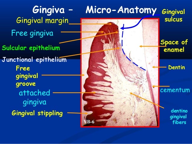
Oral Mucosa
 www.slideshare.net
mucosa gingiva epithelium gingival interdental junctional basal fibers lamina papilla
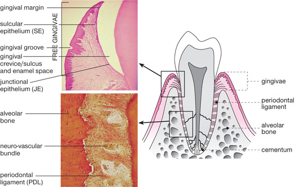
10+ Gingival Anatomy - GladeleKatelynn
 gladelekatelynn.blogspot.com
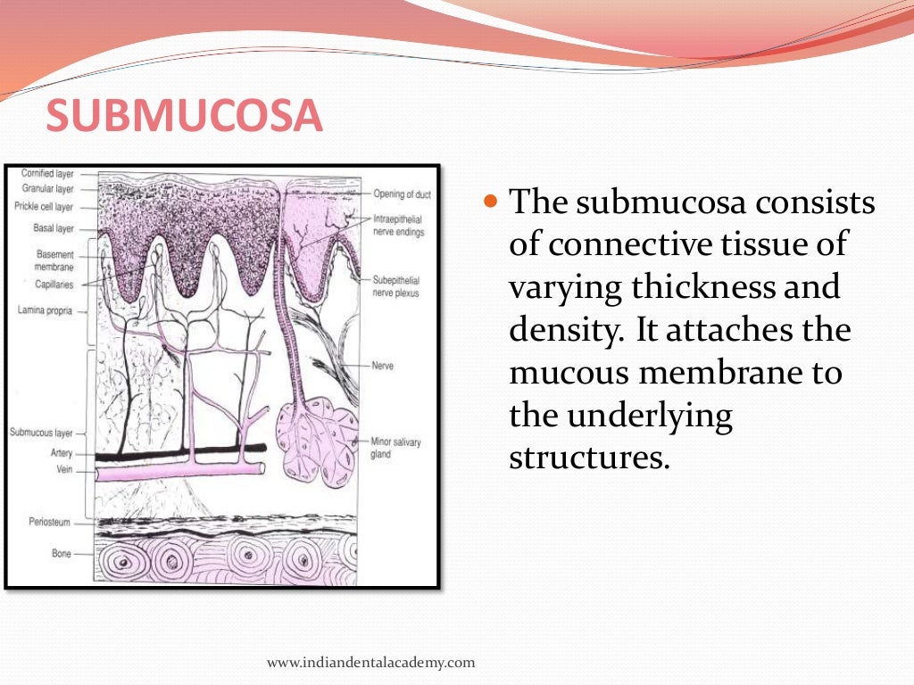
Oral Mucous Membranes-2 /certified Fixed Orthodontic Courses By India…
 www.slideshare.net
mucous submucosa orthodontic courses certified membrane membranes omm
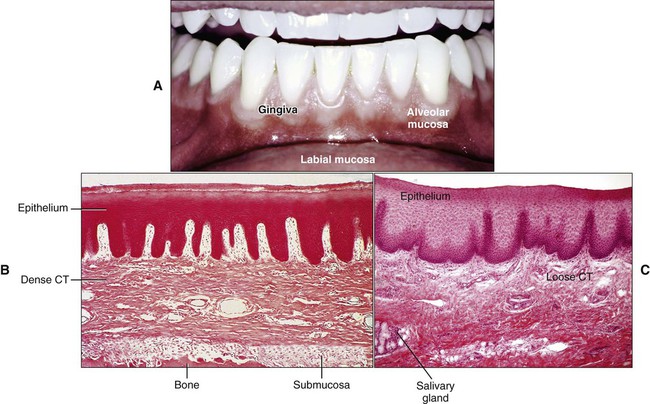
1: Structure Of The Oral Tissues | Pocket Dentistry
 pocketdentistry.com
oral mucosa tissues pocketdentistry
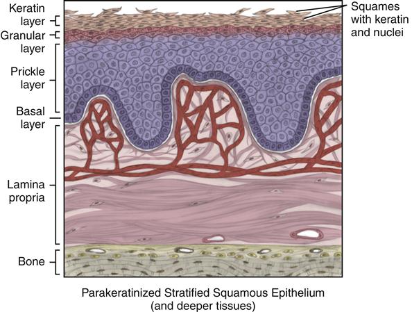
Lip Mucosa Histology | Sitelip.org
 sitelip.org
mucosa histology
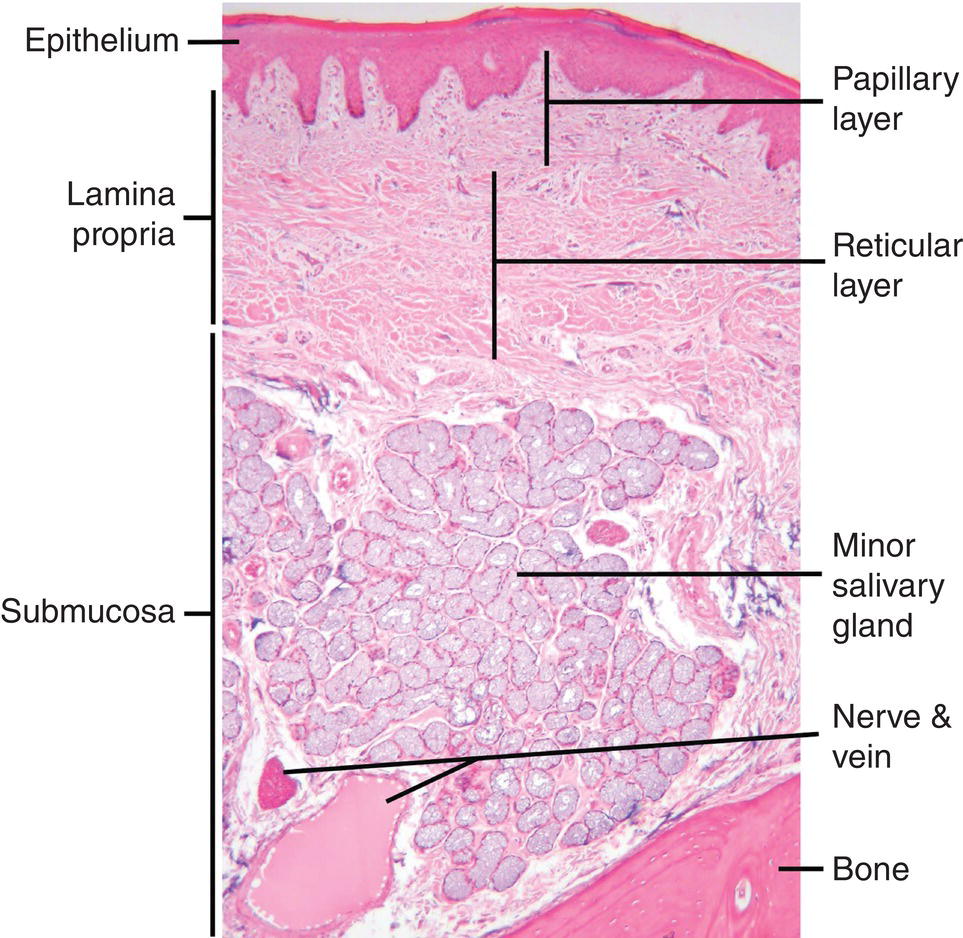
Oral Histology Digital Lab Mucosa Section Through The Gingiva Image
 www.sexiezpix.com
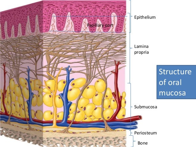
Periodontium 1
 www.slideshare.net
mucosa keratinized periodontium basal lamina propria mucous membrane epithelium periosteum epith submucosa
Oral Mucosa Part 1: Layers Of Oral Epithelium. - YouTube
 www.youtube.com
mucosa epithelium
Periodontics. - Ppt Download
 slideplayer.com
1A: Epithelial Desquamation In Alveolar Mucosa; 1B: Peeling Biopsy
 www.researchgate.net
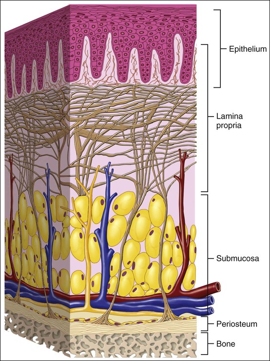
12: Oral Mucosa | Pocket Dentistry
 pocketdentistry.com
oral mucosa tissue components main figure pocketdentistry
Hypothesis For Minimally Invasive Mandibular Bone Augmentation. (a) The
 www.researchgate.net
bone periosteum mandibular augmentation mucosa invasive hypothesis minimally subperiosteal needle
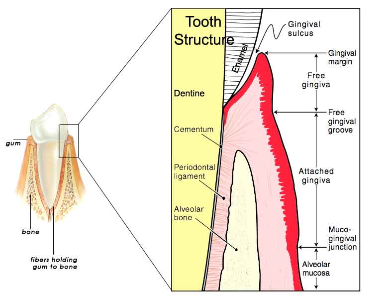
Gingival Sulcus Of The Tooth | News | Dentagama
 dentagama.com
Mucosa histology. Gingiva attached interdental papilla gingival mucosa mucogingival margin vestibular fold fornix frenum posterior. 9- oral mucosa (1) – composition and layers