← periosteum and oral mucosa Oral audie – telegraph connective tissue with the periosteum Periosteum bone anatomy tissue blood vessels nerves structure connective system meaning muscle nerve dentagama supply peri →
If you are looking for 200+ Periosteum Stock Photos, Pictures & Royalty-Free Images - iStock you've visit to the right web. We have 35 Pictures about 200+ Periosteum Stock Photos, Pictures & Royalty-Free Images - iStock like Periosteum, Pacinian Corpuscle In Periosteum Photograph by Jose Calvo / Science and also Photomicrographs of periosteum group (H & E). A. At 2 weeks, B. At. Here you go:
200+ Periosteum Stock Photos, Pictures & Royalty-Free Images - IStock
 www.istockphoto.com
Periosteum Histology
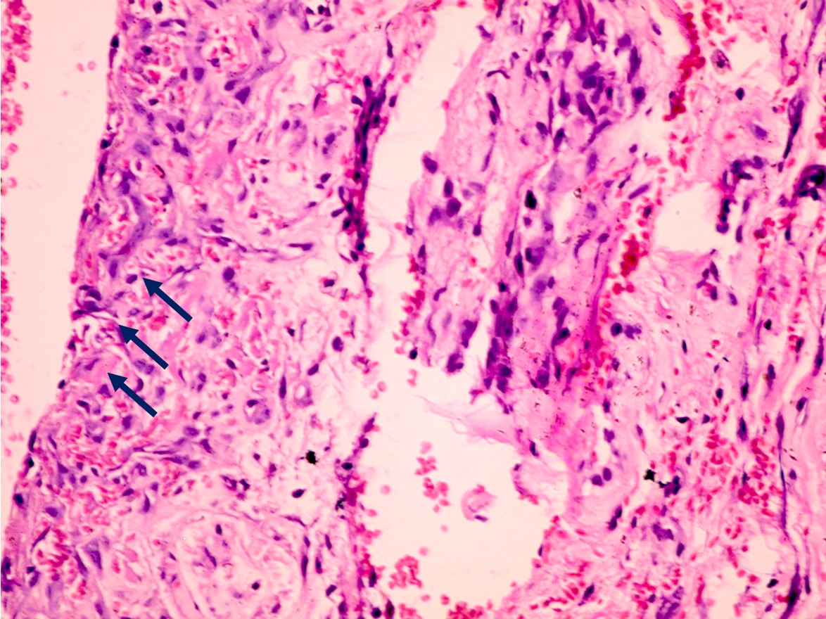 ar.inspiredpencil.com
Photomicrograph Of Compact Bone Labeled
 mavink.com
Periosteum Histology Slide Diagram | Quizlet
 quizlet.com
Histology, Periosteum And Endosteum | Treatment & Management | Point Of
 www.statpearls.com
Caption
 histology.oucreate.com
Bone Cells Under A Microscope
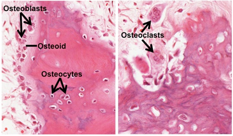 www.animalia-life.club
Histological Analysis Of Periosteum (P) And IM Samples. (A-F) Analysis
 www.researchgate.net
periosteum histological fibrous layer bv
SPONGY BONE HISTOLOGY | Microanatomy Web Atlas | Gwen V. Childs, Ph.D.
 microanatomy.net
bone histology spongy periosteum microanatomy cannon where layers osteogenic osteoblasts cells htm
Light Microscopy Images Of The Rat Tibia Periosteum Treated (b And D
 www.researchgate.net
Periosteum Histology
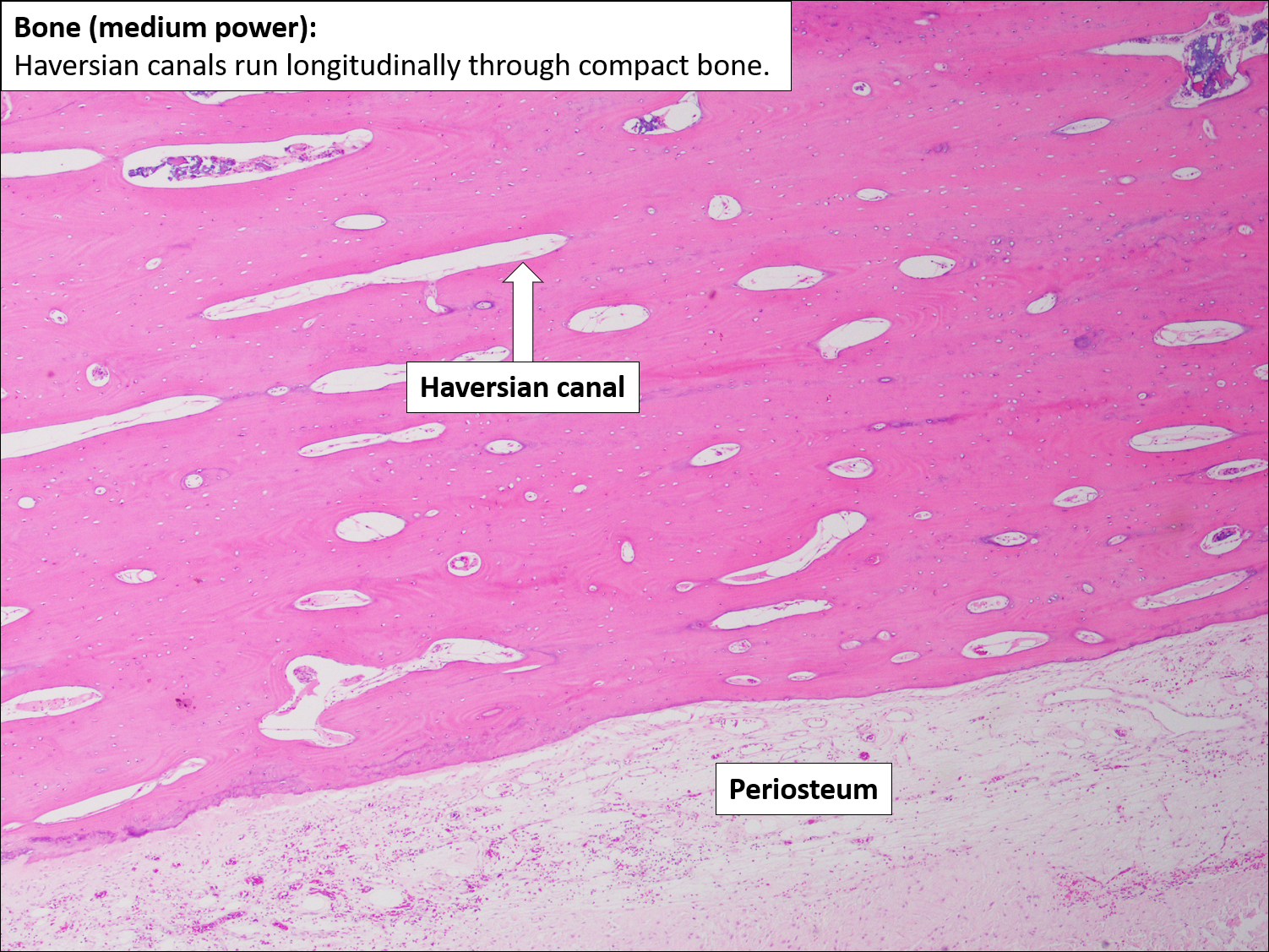 ar.inspiredpencil.com
Bone Cellsosteoblasts Osteocytes And Osteoclasts
 fity.club
A Photomicrograph Of A Longitudinal Section Of The Proximal End Of The
 www.researchgate.net
Células óseas, Sección, Micrografía De Luz 20X. Hueso Compacto Con
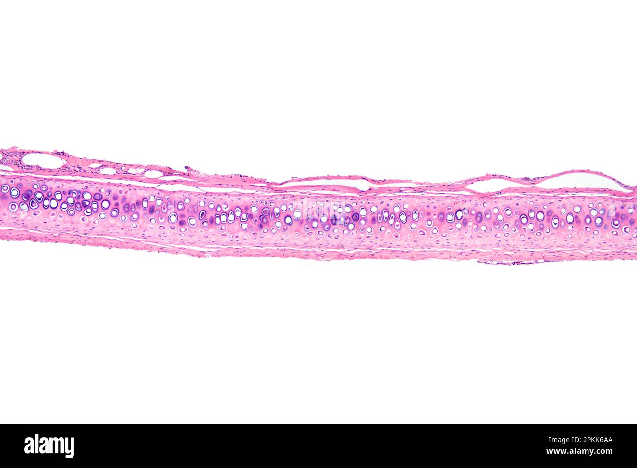 www.alamy.es
Histological Staining Of Human Periosteum Samples, Harvested Close To A
 www.researchgate.net
[PDF] The Periosteum. Part 1: Anatomy, Histology And Molecular Biology
![[PDF] The periosteum. Part 1: Anatomy, histology and molecular biology](https://d3i71xaburhd42.cloudfront.net/1f4dc57f426e15961d4df579c3f4a17eebc7461c/4-Figure1-1.png) www.semanticscholar.org
www.semanticscholar.org
Compact Bone Microscope Labeled
 anatomybrainley57.netlify.app
Bone Histology - Embryology
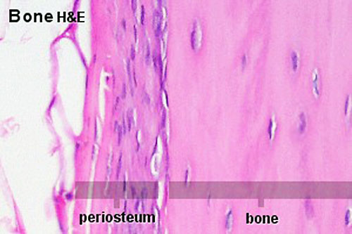 embryology.med.unsw.edu.au
bone periosteum histology endosteum embryology development au musculoskeletal
Normal Periosteum Diagram | Quizlet
 quizlet.com
Periosteum And Bone Photograph By Microscape
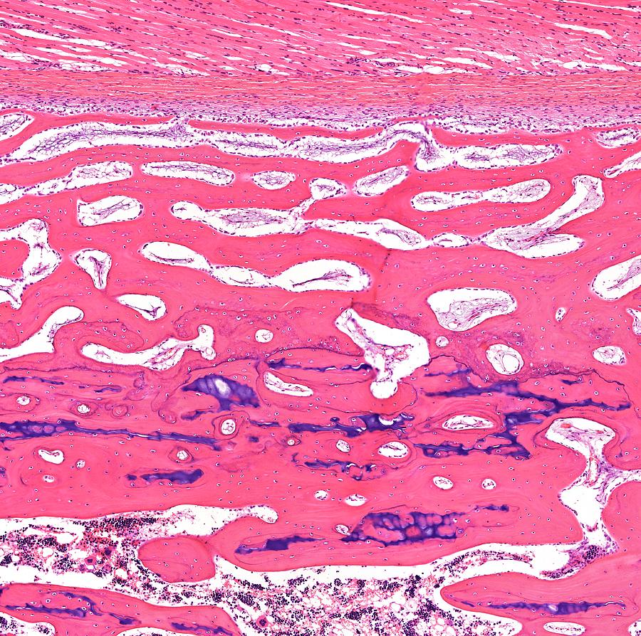 pixels.com
Micrographs Of Periosteum From (A) Control And (B) ESW-stimulated
 www.researchgate.net
Stevenel's Blue/ Van Gieson's Picrofuchsin Stained Samples For Light
 www.researchgate.net
Histology, Periosteum And Endosteum | Treatment & Management | Point Of
 www.statpearls.com
[PDF] The Periosteum. Part 1: Anatomy, Histology And Molecular Biology
![[PDF] The periosteum. Part 1: Anatomy, histology and molecular biology](https://d3i71xaburhd42.cloudfront.net/1f4dc57f426e15961d4df579c3f4a17eebc7461c/5-Figure2-1.png) www.semanticscholar.org
www.semanticscholar.org
Pacinian Corpuscle In Periosteum Photograph By Jose Calvo / Science
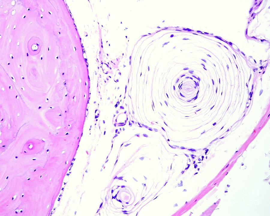 fineartamerica.com
Two Types Of Fibroblasts In The Periosteum.: (a) Typical Fibroblasts
 www.researchgate.net
fibroblasts observed fibre perforating periosteum
Intramembranous Ossification Stock Image - Image Of Histology
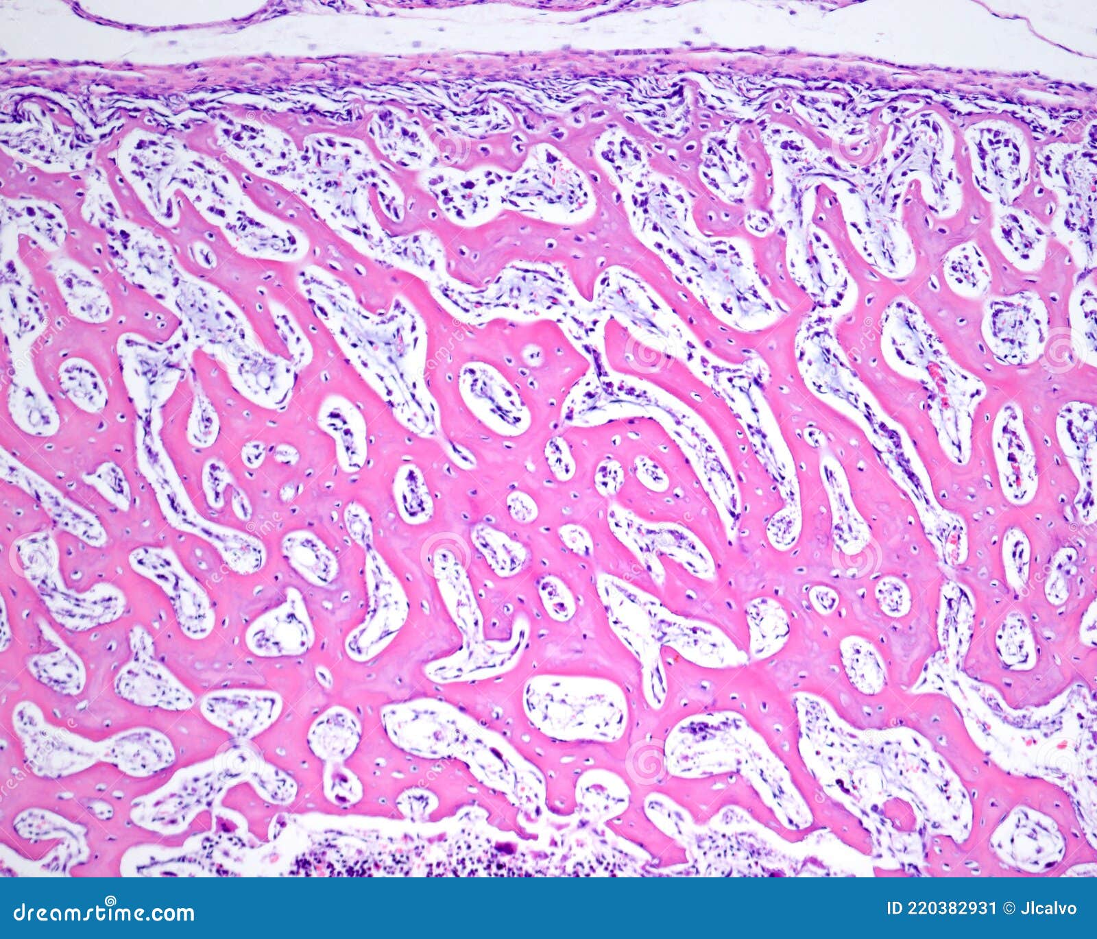 www.dreamstime.com
160+ Periosteum Stock Photos, Pictures & Royalty-Free Images - IStock
 www.istockphoto.com
Photomicrographs Of Periosteum Group (H & E). A. At 2 Weeks, B. At
 www.researchgate.net
A Photomicrograph Of Diaphysis Of A Control Rat Showing Periosteum (P
 www.researchgate.net
Periosteum Stok Fotoğraf, Resimler Ve Görseller - IStock
 www.istockphoto.com
(a) Conventional Light Microscopy. Hematoxylin And Eosin-stained
 www.researchgate.net
Periosteum
 ar.inspiredpencil.com
Developing Bone Histology
 ar.inspiredpencil.com
Cartilage Histology
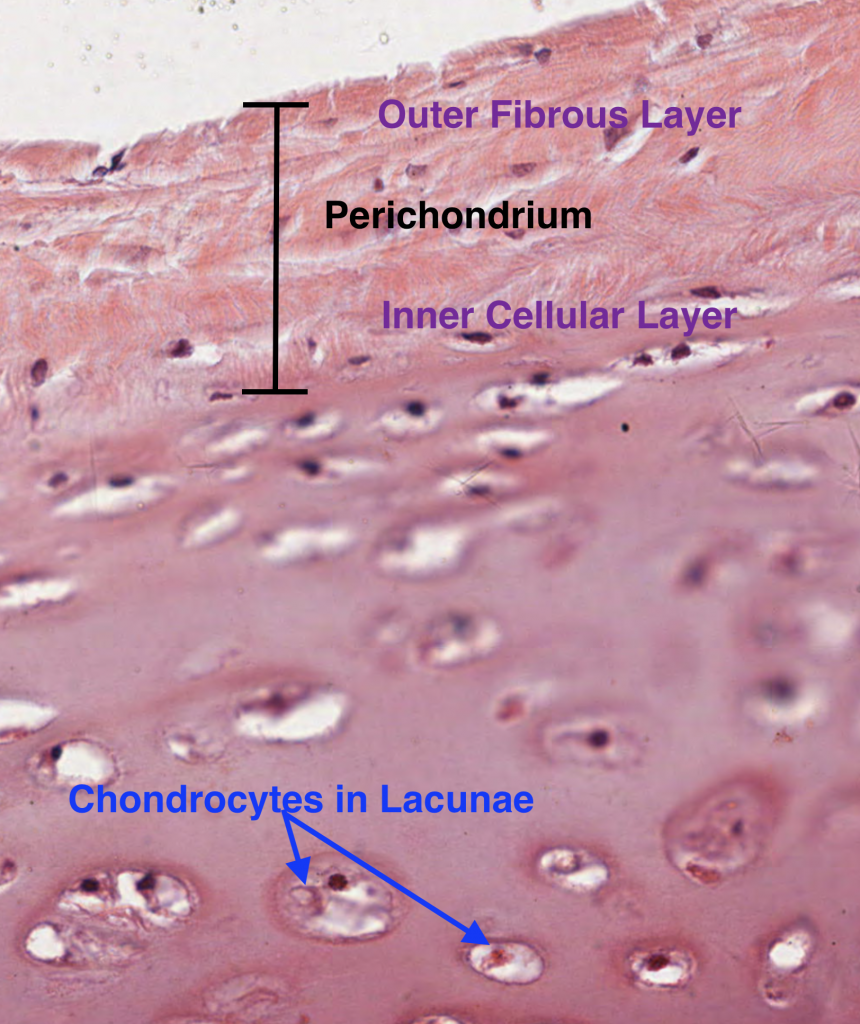 mavink.com
Fibroblasts observed fibre perforating periosteum. Histology, periosteum and endosteum. Periosteum histological fibrous layer bv